






 9PGW
9PGW
Crystal structure of STEAP2 N-domain soaked with NADP+ (second collection)
Summary for 9PGW
| Entry DOI | 10.2210/pdb9pgw/pdb |
| Descriptor | Metalloreductase STEAP2, NADPH DIHYDRO-NICOTINAMIDE-ADENINE-DINUCLEOTIDE PHOSPHATE, 1,2-ETHANEDIOL, ... (5 entities in total) |
| Functional Keywords | oxidoreductase |
| Biological source | Homo sapiens (human) |
| Total number of polymer chains | 1 |
| Total formula weight | 21616.13 |
| Authors | |
| Primary citation | Shin, I.,Sun, L.Z.,Liu, A. Structural and spectroscopic resolution of the NADPH redox state in the STEAP2 cytosolic oxidoreductase domain. J.Biol.Chem., 301:110822-110822, 2025 Cited by PubMed Abstract: The six-transmembrane epithelial antigen of prostate (STEAP) family of membrane proteins comprises four human metalloreductases essential for iron and copper homeostasis, redox balance, and cell proliferation. These enzymes transfer electrons from cytosolic NADPH to extracellular ferric and cupric ions via a FAD and heme-dependent pathway. STEAP2, 3, and four contain an N-terminal cytosolic oxidoreductase domain (OxRD) that enables electron input from NADPH, making STEAP2 ideal for studying redox-state cofactor or cosubstrate dynamics. While recent structures of STEAP proteins have been crucial, the redox state of bound NADPH has remained ambiguous in structural data, limiting mechanistic understanding. Here, we address this key missing piece of ambiguity for understanding the electron transfer pathway. We report high-resolution crystal structures of the STEAP2 OxRD with NADPH, in which the redox state of the cosubstrate is directly validated by single-crystal spectroscopy. Comparison with a recent cryoEM structure reveals conformational differences in the FAD-binding region, suggesting a plausible model in which domain reorientation between the N-terminal OxRD and C-terminal transmembrane domain (TMD) facilitates FADH loading and FAD release. These findings resolve a key ambiguity in STEAP structural biology and underscore the importance of experimental redox-state verification in structural studies of redox enzymes. PubMed: 41101505DOI: 10.1016/j.jbc.2025.110822 PDB entries with the same primary citation |
| Experimental method | X-RAY DIFFRACTION (1.46 Å) |
Structure validation
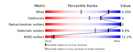
Download full validation report


