






 9OHK
9OHK
Human P2X2 receptor channel in lipid nanodiscs with 0.5mM Suramin
This is a non-PDB format compatible entry.
Summary for 9OHK
| Entry DOI | 10.2210/pdb9ohk/pdb |
| Related | 9OGH |
| EMDB information | 70496 |
| Descriptor | P2X purinoceptor 2, 2-acetamido-2-deoxy-beta-D-glucopyranose, 8,8'-[CARBONYLBIS[IMINO-3,1-PHENYLENECARBONYLIMINO(4-METHYL-3,1-PHENYLENE)CARBONYLIMINO]]BIS-1,3,5-NAPHTHALENETRISULFON IC ACID (3 entities in total) |
| Functional Keywords | atp-gated ion channel, membrane protein |
| Biological source | Homo sapiens (human) |
| Total number of polymer chains | 3 |
| Total formula weight | 161327.70 |
| Authors | Dhingra, S.,Swartz, K.J. (deposition date: 2025-05-05, release date: 2026-03-18, Last modification date: 2026-06-10) |
| Primary citation | Dhingra, S.,Moog, M.,Swartz, K.J. Mechanisms of ligand recognition and channel opening for P2X2 receptors in lipid nanodiscs. Sci Adv, 12:eaee4242-eaee4242, 2026 Cited by PubMed Abstract: Extracellular adenosine 5'-triphosphate (ATP) activates P2X receptor channels (P2XRs) that serve important roles in the immune and nervous systems. Available structures of P2XRs in detergents reveal that ATP binding to the extracellular domain leads to severing of subunit interfaces within transmembrane regions as the pore opens. Here we report cryo-electron microscopy structures of the human P2X2R in lipid nanodiscs in an apo closed state, with ATP, Mg-ATP, and suramin bound. We find that a unique Arg residue interacts with the γ-PO of ATP in P2X2R and underlies the requirement of this subtype for ATP. Channel opening and desensitization occur when ATP binds, whereas the channel remains closed when Mg-ATP binds. A continuous belt of partially resolved lipids in the outer leaflet stabilizes the closed state, and the presence of lipids prevents the severing of subunit interfaces as the channel opens. These findings establish key mechanistic principles of gating for P2X2R in a membrane-like environment, providing a framework for future mechanistic studies and therapeutic development. PubMed: 42202011DOI: 10.1126/sciadv.aee4242 PDB entries with the same primary citation |
| Experimental method | ELECTRON MICROSCOPY (3.2 Å) |
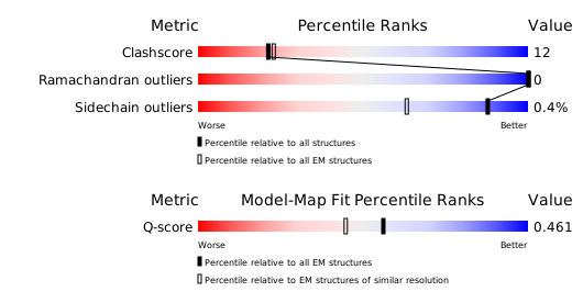
Structure validation
Download full validation report


