






 9NET
Cryo-EM of NBD-L4pY filament at 0.4 mM concentration
Summary for 9NET
| Entry DOI | 10.2210/pdb9net/pdb |
| EMDB information | 49335 |
| Descriptor | (UQ4)LLLL(PTR) (1 entity in total) |
| Functional Keywords | peptide fiber, helical polymer, protein fibril |
| Biological source | synthetic construct |
| Total number of polymer chains | 1 |
| Total formula weight | 947.97 |
| Authors | |
| Primary citation | Guo, J.,Zia, A.,Guo, W.,Tachiyama, S.,Qiu, K.,Wang, J.,Botting, J.,Liu, Z.,Qiao, Y.,Diao, J.,Liu, J.,Wang, F.,Xu, B. In Situ Multiscale Imaging Reveals Cellular Selection and Remodeling of Peptide Assemblies from Membrane Perturbation to Actin Dysregulation. J.Am.Chem.Soc., 148:40407-40418, 2026 Cited by PubMed Abstract: Enzyme-responsive peptide nanomaterials can induce non-apoptotic cancer cell death, yet the supramolecular states that mediate activity in cellular environments are usually inferred rather than directly resolved. Here, we integrate multiscale in situ imaging with biochemical assays to map distinct assembly states of the phosphatase-responsive peptide NBD-L4pY across cell-free and cellular environments. Cell-free cryo-electron microscopy (cryo-EM) reveals concentration- and history-dependent cross-β polymorphs. An equilibrated NBD-L4pY solution shows no detectable filamentous assemblies, whereas rapid dilution from a higher concentration produces a mixed filament population containing a ∼7 nm C13 nanotube. At the cell interface, this accessible C13 packing architecture becomes predominant and is resolved in situ at 3.4 Å. Cryo-electron tomography (cryo-ET) places these nanotubes to membrane contact and invagination sites associated with localized membrane perturbation and early permeability changes. At later times, cryo-ET of lamellae prepared by cryo-focused ion beam (cryo-FIB) milling reveals morphologically distinct intracellular assemblies associated with filamentous actin and membranous organelles. Fractionation shows profilin-1 (PFN1) redistribution into peptide-rich pellets, whereas reconstitution demonstrates dose-dependent impairment of actin polymerization. Together, the data support a two-stage model in which cellular environments first favor a membrane-active nanotube and subsequently promote supramolecular remodeling into morphologically distinct intracellular assemblies associated with altered actin regulation. Thus, context-dependent structure selection and remodeling, rather than enzyme activation alone, emerge as key determinants of bioactive supramolecular materials. PubMed: 42938067DOI: 10.1021/jacs.6c14737 PDB entries with the same primary citation |
| Experimental method | ELECTRON MICROSCOPY (3.5 Å) |
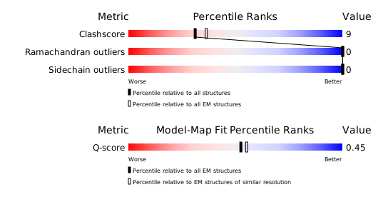
Structure validation
Download full validation report


