






 9MRX
9MRX
Cryo-EM structure of KwaA-KwaB complex
Summary for 9MRX
| Entry DOI | 10.2210/pdb9mrx/pdb |
| EMDB information | 48564 |
| Descriptor | Kiwa protein KwaA, Kiwa protein KwaB (2 entities in total) |
| Functional Keywords | kwaa-kwab complex, anti-phage defense, immune system |
| Biological source | Escherichia coli More |
| Total number of polymer chains | 11 |
| Total formula weight | 343138.53 |
| Authors | Zhang, Z.,Patel, D.J. (deposition date: 2025-01-08, release date: 2025-08-06, Last modification date: 2026-05-20) |
| Primary citation | Zhang, Z.,Todeschini, T.C.,Wu, Y.,Kogay, R.,Naji, A.,Cardenas Rodriguez, J.,Mondi, R.,Kaganovich, D.,Taylor, D.W.,Bravo, J.P.K.,Teplova, M.,Amen, T.,Koonin, E.V.,Patel, D.J.,Nobrega, F.L. Kiwa is a membrane-embedded defense supercomplex activated at phage attachment sites. Cell, 188:5862-5877.e23, 2025 Cited by PubMed Abstract: Bacteria and archaea deploy diverse antiviral defense systems, many of which remain mechanistically uncharacterized. Here, we characterize Kiwa, a widespread two-component system composed of the transmembrane sensor KwaA and the DNA-binding effector KwaB. Cryogenic electron microscopy (cryo-EM) analysis reveals that KwaA and KwaB assemble into a large, membrane-associated supercomplex. Upon phage binding, KwaA senses infection at the membrane, leading to KwaB binding of ejected phage DNA and inhibition of replication and late transcription, without inducing host cell death. Although KwaB can bind DNA independently, its antiviral activity requires association with KwaA, suggesting spatial or conformational regulation. We show that the phage-encoded DNA-mimic protein Gam directly binds and inhibits KwaB but that co-expression with the Gam-targeted RecBCD system restores protection by Kiwa. Our findings support a model in which Kiwa coordinates membrane-associated detection of phage infection with downstream DNA binding by its effector, forming a spatially coordinated antiviral mechanism. PubMed: 40730155DOI: 10.1016/j.cell.2025.07.002 PDB entries with the same primary citation |
| Experimental method | ELECTRON MICROSCOPY (3.75 Å) |
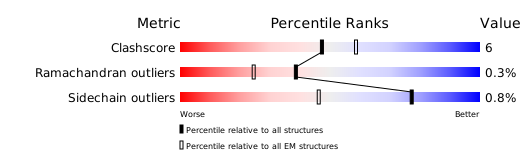
Structure validation
Download full validation report


