Loading
PDBj
MenuPDBj@FacebookPDBj@X(formerly Twitter)PDBj@BlueSkyPDBj@YouTubewwPDB FoundationwwPDBDonate
RCSB PDBPDBeBMRBAdv. SearchSearch help

9MRO

Crystal structures of a cyanobacterial DAP epimerase bound to either D,L-aziDAP or D,L-alpha-methyl DAP

Summary for 9MRO
Entry DOI10.2210/pdb9mro/pdb
DescriptorDiaminopimelate epimerase, (2R,6S)-2,6-DIAMINO-2-METHYLHEPTANEDIOIC ACID (3 entities in total)
Functional Keywordsdap epimerase, diaminopimelic acid, dapf, cofactor-independent, protein biochemistry, enzyme mechanism, inhibitor, antibiotic, lysine biosynthesis
Biological sourceAnabaena sp. YBS01
Total number of polymer chains4
Total formula weight125530.91
Authors
Chen, P.,Lamer, T.,Vederas, J.C.,Lemieux, M.J. (deposition date: 2025-01-08, release date: 2025-07-16, Last modification date: 2026-07-29)
Primary citationLamer, T.,Chen, P.,Catenza, K.,Perov, I.,L, B.,Hsiao, Y.T.,Van Oers, T.J.,Lemieux, M.J.,Vederas, J.C.
Structure and inhibition of diaminopimelic acid epimerase by slow-binding alpha-methyl amino acids.
Protein Sci., 34:e70139-e70139, 2025
Cited by
PubMed Abstract: Cofactor-independent racemases and epimerases produce D-amino acids from their L-isomers for a variety of biological processes. These enzymes operate via an unusual mechanism that relies on an active site cysteine thiolate (pK ~ 8.5) to deprotonate an amino acid α-carbon (pK ~ 29) and are of interest not only because of their biocatalytic potential for D-amino acid production, but also because many play key roles in biology and are antibiotic targets. However, obtaining crystal structures of these enzymes, especially in their closed, substrate- or inhibitor-bound conformations, is difficult. In this work, we characterized diaminopimelic acid (DAP) epimerase from the cyanobacterium Anabaena. DAP epimerase has long been of interest as an antibiotic target as it converts L,L-DAP to D,L-DAP for lysine and peptidoglycan biosynthesis. We solved three crystal structures of this enzyme in its closed, inhibitor-bound conformation, up to a resolution of 1.5 Å. Two structures show the enzyme covalently bound through its catalytic cysteine residues to previously reported aziridine-based inhibitors. One structure unexpectedly shows the enzyme bound to a different compound, D,L-α-methylDAP, presumably produced as a synthetic byproduct. Stereoselective synthesis of L,L- and D,L-α-methylDAP followed by inhibition assays shows that these compounds are slow-binding inhibitors of DAP epimerase. α-MethylDAP inhibitors provide a more accessible alternative to aziridine-based inhibitors to obtain crystal structures of DAP epimerase in its closed conformation. Comparisons of bacterial, cyanobacterial, and plant DAP epimerases provided here offer new insights into functional and structural differences between these enzymes.
PubMed: 40299312
DOI: 10.1002/pro.70139
PDB entries with the same primary citation
Experimental method
X-RAY DIFFRACTION (1.59 Å)
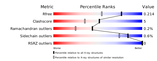
Structure validation

259351

PDB entries from 2026-09-09

PDB statisticsPDBj update infoContact PDBjnumon