






 9LNU
9LNU
Structure of MBP tagged H2A.W-H2B
Summary for 9LNU
| Entry DOI | 10.2210/pdb9lnu/pdb |
| Descriptor | Structure of MBP tagged H2A.W-H2B, SULFATE ION (3 entities in total) |
| Functional Keywords | h2a-h2b, h3-h4, histone variant, nucleosome, h2a.w, nuclear protein |
| Biological source | Arabidopsis thaliana |
| Total number of polymer chains | 1 |
| Total formula weight | 65612.14 |
| Authors | |
| Primary citation | Hu, S.,Yang, Y.,Wang, L.,Xu, L. Crystal structure of the histone heterodimer containing histone variant H2A.W. Biochem Biophys Rep, 46:102535-102535, 2026 Cited by PubMed Abstract: Histone variant H2A.W plays a critical role in heterochromatin organization and genome stability, the loss of H2A.W can lead to heterochromatin decondensation, resulting in growth defects in flowering plants. Yet the mechanism underlying how H2A.W integrates into chromatin remains elusive. Here, we present the first high-resolution crystal structure of the H2A.W (AtH2A.W) - H2B (HsH2B) in the DNA-free heterodimeric state. In the structure, the global domain of DNA-free AtH2A.W-HsH2B share almost the same structure with its DNA bound form in the nucleosome, except the αC helix and the following C-terminal tail region (docking domain). What's more, we find that the AtH2A.W docking domain binds to the α2-α3 region of HsH2B, which is different from its role of interact with H3-H4 and DNA in nucleosome. These structure analyses suggest that the αC and docking domain of AtH2A.W is highly dynamic and may be remodeled during nucleosome assembly. In summary, our findings highlight the dynamic nature of AtH2A.W docking domain, providing mechanistic insight into how AtH2A.W integrates into chromatin and supports specialized chromatin functions. PubMed: 41847302DOI: 10.1016/j.bbrep.2026.102535 PDB entries with the same primary citation |
| Experimental method | X-RAY DIFFRACTION (1.85 Å) |
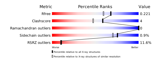
Structure validation
Download full validation report


