






 9KY5
9KY5
Structure of y+LAT2 bound with Arg
Summary for 9KY5
| Entry DOI | 10.2210/pdb9ky5/pdb |
| EMDB information | 62640 |
| Descriptor | Y+L amino acid transporter 2, Amino acid transporter heavy chain SLC3A2, ARGININE (3 entities in total) |
| Functional Keywords | complex, membrane protein, amino acid |
| Biological source | Homo sapiens (human) More |
| Total number of polymer chains | 2 |
| Total formula weight | 128946.83 |
| Authors | |
| Primary citation | Dai, L.,Xu, K.,Zhang, T.,Wang, X.,Zeng, Q.,Liang, H.,Xu, C.,Yang, L.,Wang, Z.,Yan, R. Structural insights into the human system y + L amino acid transporter complex. Structure, 33:1224-1232.e5, 2025 Cited by PubMed Abstract: System yL facilitates the sodium-independent transport of cationic and sodium-dependent transport of neutral amino acids via heteromeric amino acid transporters. System yL consists of either SLC7A6 (yLAT2) or SLC7A7 (yLAT1) and 4F2hc (SLC3A2). The yLAT2-4F2hc complex mediates the exchange of -lysine (Lys), -arginine (Arg), -leucine (Leu), and -glutamine (Gln) and is important for the glutamate-glutamine cycle and ammonia clearance. c-Myc-driven upregulation of yLAT2 in cancer enhances amino acid uptake and mTORC1 activation, promoting tumor growth. Its transport mechanism has remained unclear. Here, we determined the cryoelectron microscopic (cryo-EM) structures of the yLAT2-4F2hc complex bound to either Arg or Leu at 3.60 Å and 3.58 Å resolution, respectively, revealing an outward-open conformation. Our structural analysis highlights conformational changes during transport, and functional assays validate critical residues involved in substrate binding and transport. These findings elucidate the molecular mechanism of the system yL and provide a foundation for developing targeted therapies against yLAT2. PubMed: 40347950DOI: 10.1016/j.str.2025.04.012 PDB entries with the same primary citation |
| Experimental method | ELECTRON MICROSCOPY (3.6 Å) |
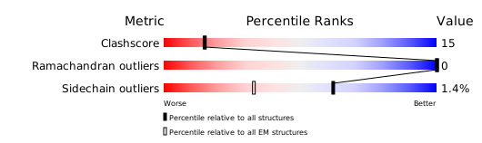
Structure validation
Download full validation report


