






 9JTE
9JTE
Crystal structure of SARS-CoV-2 receptor binding domain complexed with fox ACE2
Summary for 9JTE
| Entry DOI | 10.2210/pdb9jte/pdb |
| Descriptor | Angiotensin-converting enzyme, Spike protein S1, beta-D-mannopyranose-(1-4)-2-acetamido-2-deoxy-beta-D-glucopyranose-(1-4)-2-acetamido-2-deoxy-beta-D-glucopyranose, ... (5 entities in total) |
| Functional Keywords | sars-cov-2, receptor binding domain, fox ace2, viral protein |
| Biological source | Vulpes vulpes (red fox) More |
| Total number of polymer chains | 4 |
| Total formula weight | 184362.29 |
| Authors | Jun, L.,Xiaoyan, N. (deposition date: 2024-10-04, release date: 2025-10-22, Last modification date: 2026-05-06) |
| Primary citation | Wang, C.,Nan, X.,Pei, C.,Li, M.,Wu, J.,Wang, Z.,Fan, S.,Lan, J. Molecular basis of fox ACE2 recognition by receptor binding domains of SARS-CoV-2 and PCoV-GD. Cell Insight, 5:100314-100314, 2026 Cited by PubMed Abstract: The severe acute respiratory syndrome coronavirus 2 (SARS-CoV-2) positive RNA detected in red fox swab samples provide evidence of a natural SARS-CoV-2 infection in this species. Many studies have also demonstrated that the red fox angiotensin converting enzyme 2 (fACE2) could serve as the receptor of SARS-CoV-2 and many SARS-CoV-2 related sarbecoviruses. However, the molecular mechanisms of fACE2 attached by animal-origin coronaviruses remain poorly understood. Here, we found that fACE2 could mediate pseudovirus entry and cell membrane fusion induced by the SARS-CoV-2 and pangolin coronavirus isolated from Guangdong (PCoV-GD) spikes as human ACE2 (hACE2). The binding affinity of fACE2 bound to the receptor-binding domain (RBD) of PCoV-GD was stronger than the SARS-CoV-2 RBD demonstrated by surface plasmon resonance (SPR) assay, while deglycosylation of PCoV-GD RBD N370 by T372A mutation or glycosylation of SARS-CoV-2 RBD N370 by A372T mutation had little effect on fACE2 binding compared with wild-type (WT) RBD. We further solved the crystal structures of fACE2 bound to SARS-CoV-2 RBD and PCoV-GD T372A mutant RBD. Interface analysis and SPR assay revealed that the R417 and H498 of PCoV-GD RBD might account for the binding affinity enhancement with fACE2 compared with K417 and Q498 of SARS-CoV-2 RBD. Moreover, the Y453F mutation in SARS-CoV-2 RBD increased the binding affinity to fACE2 while this mutation in PCoV-GD RBD decreased the binding affinity to fACE2 compared with the corresponding WT RBDs. Furthermore, the RBDs of many prevalent SARS-CoV-2 variants could all bind to fACE2. Our results indicated that the continuous surveillance of SARS-CoV-2 and related sarbecoviruses in fox species was also necessary to better prevent animal-to-human spillover of the coronaviruses. PubMed: 41960421DOI: 10.1016/j.cellin.2026.100314 PDB entries with the same primary citation |
| Experimental method | X-RAY DIFFRACTION (3.66 Å) |
Structure validation
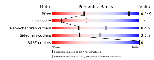
Download full validation report


