






 9JHQ
9JHQ
Crystal structure of GodF, a post-translational modification enzyme involved in the biosynthesis of goadsporin
Summary for 9JHQ
| Entry DOI | 10.2210/pdb9jhq/pdb |
| Descriptor | Goadsporin biosynthetic protein, ACETATE ION, GLYCEROL, ... (4 entities in total) |
| Functional Keywords | trna-dependent glutamylation, biosynthetic protein, goadsporin, transferase |
| Biological source | Streptomyces sp. TP-A0584 |
| Total number of polymer chains | 1 |
| Total formula weight | 93965.97 |
| Authors | Shimizu-Ibuka, A.,Kato, Y. (deposition date: 2024-09-10, release date: 2025-09-10, Last modification date: 2026-03-04) |
| Primary citation | Shimizu-Ibuka, A.,Kato, Y.,Asamizu, S.,Onaka, H. Structural Analysis of GodF, an O-glutamylation Enzyme Involved in Goadsporin Biosynthesis. J.Mol.Biol., 438:169619-169619, 2026 Cited by PubMed Abstract: Goadsporin is one of linear azole-containing peptides (LAPs) that form a subgroup within ribosomally synthesized and post-translationally modified peptides (RiPPs). It contains two dehydroalanine residues formed through the action of two enzymes, GodF and GodG, in a two-step process involving serine O-glutamylation followed by elimination. Here, we report the X-ray crystal structure of GodF, which catalyzes the tRNA-dependent glutamylation of target serine residues, resolved at a 2.34-Å resolution. Although GodF exhibits low homology at the primary sequence level, its overall structure closely resembles that of TbtB, a tRNA-dependent enzyme involved in thiopeptide biosynthesis, as well as the O-glutamylation domains of NisB and MibB, which serve as dehydroalanine synthases in lanthipeptide biosynthesis. The residues and structural elements forming the active site are well-aligned among these enzymes, while regions outside the active site are poorly conserved. Like TbtB, GodF features a coiled-coil subdomain at its N-terminus, and AlphaFold3 predicts this region plays a key role in recognizing the substrate tRNA. GodF also contains a typical RiPP recognition element (RRE) motif; however, the spatial arrangement of the secondary structural elements comprising this motif differs notably from those in other O-glutamylating enzymes. These structural characteristics of GodF highlight the diversity of substrate-binding pockets among RiPP-modifying enzymes, reflecting the variability in their substrate peptides and the necessity to accommodate distinct conformational and physicochemical properties. PubMed: 41478604DOI: 10.1016/j.jmb.2025.169619 PDB entries with the same primary citation |
| Experimental method | X-RAY DIFFRACTION (2.34 Å) |
Structure validation
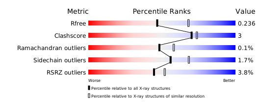
Download full validation report


