






 9IJO
9IJO
X-ray crystal structure of F46C myoglobin with a covalently linked 4-methyl-2,2'-bipyridine group in complex with Cu2+
This is a non-PDB format compatible entry.
Summary for 9IJO
| Entry DOI | 10.2210/pdb9ijo/pdb |
| Descriptor | Myoglobin, PROTOPORPHYRIN IX CONTAINING FE, 4-methyl-2-(4-methylpyridin-2-yl)pyridine, ... (5 entities in total) |
| Functional Keywords | heme protein, myoglobin, copper binding, metal binding protein |
| Biological source | Physeter catodon (sperm whale) |
| Total number of polymer chains | 1 |
| Total formula weight | 18186.39 |
| Authors | Lin, Y.W. (deposition date: 2024-06-25, release date: 2025-07-02, Last modification date: 2026-08-12) |
| Primary citation | Nie, L.S.,Liu, X.C.,Yu, L.,Liu, A.K.,Sun, L.J.,Gao, S.Q.,Lin, Y.W. Rational Design of an Artificial Metalloenzyme by Constructing a Metal-Binding Site Close to the Heme Cofactor in Myoglobin. Inorg.Chem., 63:18531-18535, 2024 Cited by PubMed Abstract: In this study, we constructed a metal-binding site close to the heme cofactor in myoglobin (Mb) by covalently attaching a nonnative metal-binding ligand of bipyridine to Cys46 through the F46C mutation in the heme distal site. The X-ray structure of the designed enzyme, termed F46C-mBpy Mb, was solved in the Cu(II)-bound form, which revealed the formation of a heterodinuclear center of Cu-His-HO-heme. Cu(II)-F46C-mBpy Mb exhibits not only nitrite reductase reactivity but also cascade reaction activity involving both hydrolysis and oxidation. Furthermore, F46C-mBpy Mb displays Mn-peroxidase activity by the oxidation of Mn to Mn using HO as an oxidant. This study shows that the construction of a nonnative metal-binding site close to the heme cofactor is a convenient approach to creating an artificial metalloenzyme with a heterodinuclear center that confers multiple functions. PubMed: 39311200DOI: 10.1021/acs.inorgchem.4c03093 PDB entries with the same primary citation |
| Experimental method | X-RAY DIFFRACTION (1.53 Å) |
Structure validation
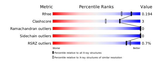
Download full validation report


