






 9E4J
9E4J
Human ASIC1a at pH 6.5 in complex with MitTx
Summary for 9E4J
| Entry DOI | 10.2210/pdb9e4j/pdb |
| EMDB information | 47512 |
| Descriptor | Acid-sensing ion channel 1, Kunitz-type neurotoxin MitTx-alpha, Basic phospholipase A2 homolog MitTx-beta (3 entities in total) |
| Functional Keywords | ion channel, trimer, toxin, membrane protein |
| Biological source | Homo sapiens (human) More |
| Total number of polymer chains | 9 |
| Total formula weight | 243414.74 |
| Authors | Hartfield, K.A.,Cahill, J.,Baconguis, I. (deposition date: 2024-10-24, release date: 2026-01-21, Last modification date: 2026-08-26) |
| Primary citation | Cahill, J.,Hartfield, K.A.,Heusser, S.A.,Ritter, N.,Poulsen, M.H.,Yoshioka, C.,Pless, S.A.,Baconguis, I. Conformational plasticity of human acid-sensing ion channel 1a. Nat.Struct.Mol.Biol., 33:1171-1182, 2026 Cited by PubMed Abstract: Acid-sensing ion channels (ASICs) are typically activated by acidic environments and contribute to nociception and synaptic plasticity. ASIC1a is the most abundant subunit in the central nervous system and forms homomeric channels permeable to Na and Ca, making it a compelling therapeutic target for acidotic pathologies including stroke and traumatic brain injury. However, a complete conformational library of human ASIC1a has yet to be described. Here we show that human ASIC1a adopts six major conformations, resolved by cryo-electron microscopy across a pH range between 8.5 and 5.7 and in the presence of a toxin agonist and a gating-modifying amino acid substitution. These major conformations establish linear transmembrane helices to be associated with an open state, delineate mechanistic differences between proton and toxin activation and demonstrate that desensitization involves unexpected conformational diversity in the transmembrane domain. Together, they provide a three-dimensional framework to integrate previous structure-function studies on ASIC. PubMed: 42463865DOI: 10.1038/s41594-026-01845-0 PDB entries with the same primary citation |
| Experimental method | ELECTRON MICROSCOPY (2.87 Å) |
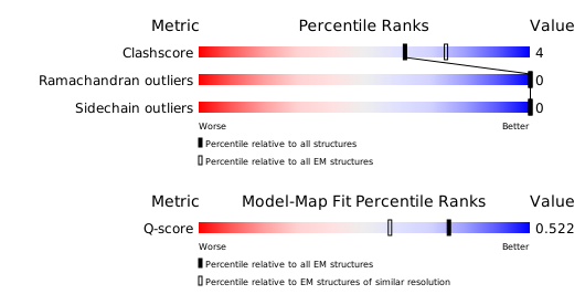
Structure validation
Download full validation report


