






 9DFS
9DFS
Structure of novel Myo7a-N isoform (ADP-bound) expressed in sensory hair cells (head domain + first two IQ domains), bound to F-actin
Summary for 9DFS
| Entry DOI | 10.2210/pdb9dfs/pdb |
| EMDB information | 46818 |
| Descriptor | Actin, alpha skeletal muscle, Unconventional myosin-VIIa, ADENOSINE-5'-DIPHOSPHATE, ... (4 entities in total) |
| Functional Keywords | actomyosin, hair cells, motor protein |
| Biological source | Oryctolagus cuniculus (rabbit) More |
| Total number of polymer chains | 4 |
| Total formula weight | 250944.83 |
| Authors | Egelman, E.H.,Shin, J.B. (deposition date: 2024-08-30, release date: 2025-09-10, Last modification date: 2026-09-23) |
| Primary citation | Li, S.,Park, J.,Phan, T.M.,Caprara, G.A.,Sittipongpittaya, N.,Sheynkman, G.M.,Peng, A.W.,Egelman, E.H.,Bird, J.E.,Shin, J.B. Tonotopic specialization of MYO7A isoforms in auditory hair cells. Nat Commun, 17:-, 2026 Cited by PubMed Abstract: Mutations in Myo7a cause Usher syndrome type 1B and non-syndromic deafness, but the precise function of MYO7A in sensory hair cells remains unclear. Using long-read sequencing, we identify and characterize a novel isoform, MYO7A-N, expressed in auditory hair cells alongside the canonical MYO7A-C. Isoform-specific knock-in mouse models reveal that inner hair cells primarily express MYO7A-C, while outer hair cells express both isoforms in opposing tonotopic gradients. Both isoforms are localized to the upper tip-link insertion site, consistent with a role in the tip link for mechanotransduction. Loss of MYO7A-N leads to outer hair cell degeneration and progressive hearing loss. Cryo-EM structures reveal isoform-specific differences at actomyosin interfaces, correlating with distinct ATPase activities. These findings reveal an unexpected layer of molecular diversity within the mechanotransduction machinery. We propose that MYO7A isoform specialization enables fine-tuning of tip-link tension, thus hearing sensitivity, and contributes to the frequency-resolving power of the cochlea. PubMed: 42236700DOI: 10.1038/s41467-026-73220-y PDB entries with the same primary citation |
| Experimental method | ELECTRON MICROSCOPY (3.1 Å) |
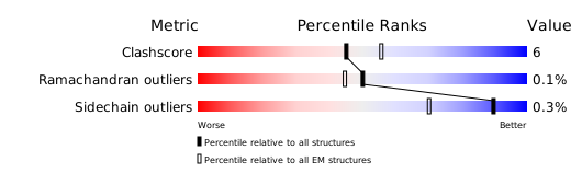
Structure validation
Download full validation report


