






 9VXW
9VXW
Cryo-EM structure of hAQP11 in LMNG
Summary for 9VXW
| Entry DOI | 10.2210/pdb9vxw/pdb |
| EMDB information | 65443 |
| Descriptor | Aquaporin-11,sfGFP, DECANE (2 entities in total) |
| Functional Keywords | membrane protein |
| Biological source | Homo sapiens (human) More |
| Total number of polymer chains | 1 |
| Total formula weight | 60153.33 |
| Authors | Suzuki, S.,Nishikawa, K.,Kamegawa, A.,kozai, D.,Fujiyoshi, Y. (deposition date: 2025-07-20, release date: 2025-12-31, Last modification date: 2026-07-15) |
| Primary citation | Suzuki, S.,Kamegawa, A.,Kozai, D.,Nishikawa, K.,Irie, K.,Fujiyoshi, Y. Cryo-EM structure of human AQP11 reveals a trimeric architecture with a large pore. Sci Adv, 12:eaeb5769-eaeb5769, 2026 Cited by PubMed Abstract: Aquaporin-11 (AQP-11) is an endoplasmic reticulum-localized water channel essential for renal development. Its structure and the molecular basis of its transport properties remained unknown. We analyzed the human AQP11 structure under cryo-electron microscopy at 2.3 Å resolution, revealing a trimeric architecture compared with other known tetrameric AQPs and a topology comprising seven transmembrane helices (Hs), including an additional N-terminal helix (H0). The channel pore is broader and more hydrophobic than that of canonical AQPs, and features a unique structure surrounding an Asn-Pro-Cys (NPC) sequence instead of the typical Asn-Pro-Ala (NPA) motif. These features provide a structural framework through which water and other small solutes can permeate AQP11. Our findings provide a blueprint for designing specific inhibitors to investigate the physiologic functions of AQP11. PubMed: 41616064DOI: 10.1126/sciadv.aeb5769 PDB entries with the same primary citation |
| Experimental method | ELECTRON MICROSCOPY (2.3 Å) |
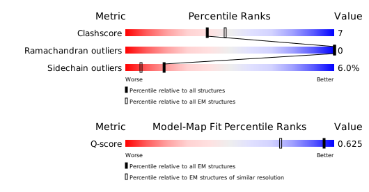
Structure validation
Download full validation report


