






 8Z4H
8Z4H
Pseudomurein Endoisopeptidase PeiR
Summary for 8Z4H
| Entry DOI | 10.2210/pdb8z4h/pdb |
| Descriptor | Endoisopeptidase PeiR (2 entities in total) |
| Functional Keywords | methanogenic archaea, pseudomurein, endopeptidases, hydrolase |
| Biological source | Methanobrevibacter ruminantium M1 |
| Total number of polymer chains | 4 |
| Total formula weight | 105085.16 |
| Authors | Guo, L.Z.,Wang, S.X.,Bai, L.p. (deposition date: 2024-04-17, release date: 2025-04-23, Last modification date: 2026-06-24) |
| Primary citation | Guo, L.,Leng, H.,Zhou, X.,Wang, S.,Bai, L. Structural and functional characterization of C39 endoisopeptidases from methanogenic archaeal phages with activity on synthetic pseudopeptidoglycan analogues. Int.J.Biol.Macromol., 367:152647-152647, 2026 Cited by PubMed Abstract: The inherent resistance of pseudopeptidoglycan to lysozyme and antibiotics has drawn considerable research attention to archaeal cell wall hydrolases from methanogenic archaeal phages. The C39 peptidases derived from methanogenic archaeal phages exhibit cleavage activity toward synthetic isopeptide substrates that mimic pseudopeptidoglycan cross-links, distinguishing them functionally from bacterial C39 peptidases, which process double glycine motifs in the ABC transport system I. However, the precise cleavage mechanism remains unclear. In this study, we systematically characterized the substrate specificities and catalytic properties of three phage-derived C39 peptidases-C39-PeiR, C39-PhiF1, and C39-PhiF3-using synthetic isopeptides and p-nitroaniline-modified peptides. X-ray crystallography and site-directed mutagenesis assays further revealed that the loop region immediately adjacent to α-helix 7 in C39-PeiR constitutes a critical structural determinant distinguishing its functional repertoire from bacterial orthologs. Additionally, C39 peptidases were classified into 16 distinct clades based on structural variations, leading to the identification of novel pseudopeptidoglycan endoisopeptidases. PubMed: 42173210DOI: 10.1016/j.ijbiomac.2026.152647 PDB entries with the same primary citation |
| Experimental method | X-RAY DIFFRACTION (2 Å) |
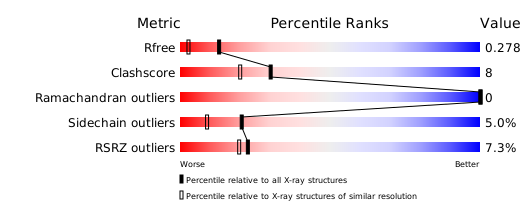
Structure validation
Download full validation report


