






 8XTU
8XTU
Crystal Structure of Rab5b GTPase domain from Leishmania donovani in complex with GDP
Summary for 8XTU
| Entry DOI | 10.2210/pdb8xtu/pdb |
| Descriptor | Rab5, MAGNESIUM ION, GUANOSINE-5'-DIPHOSPHATE, ... (4 entities in total) |
| Functional Keywords | gtpase, early endocytosis, nucleotide exchange, endocytosis |
| Biological source | Leishmania donovani |
| Total number of polymer chains | 1 |
| Total formula weight | 18945.25 |
| Authors | Zohib, M.,Pal, R.K.,Biswal, B.K.,Arora, A. (deposition date: 2024-01-11, release date: 2025-01-15, Last modification date: 2026-07-29) |
| Primary citation | Pandey, D.,Zohib, M.,Chaurasia, A.,Ansari, H.,Chowdhury, A.,Pal, R.K.,Tripathi, S.,Jain, A.,Biswal, B.K.,Siddiqi, M.I.,Arora, A. Analysis of structure and stability of Leishmania donovani Rab5a and Rab5b. Int.J.Biol.Macromol., 365:152349-152349, 2026 Cited by PubMed Abstract: Leishmania donovani Rab5 isoforms, LdRab5a and LdRab5b, play distinct roles in the early stages of endocytosis, with LdRab5a primarily regulating fluid-phase uptake and LdRab5b modulating receptor-mediated endocytosis. These functional differences are governed by structural variations within their GTPase domains. Here, we report the crystal structure of the GTPase domain of LdRab5a in its active, GppNHp-bound, form and compare it to the previously solved GDP-bound structure. Binding of the γ-phosphate analog induces a closed conformation of the Switch I and Switch II regions, a characteristic of the active state. Circular Dichroism and thermal stability assessments using Differential Scanning Calorimetry confirmed proper folding of the protein and revealed that guanine nucleotide and Mg binding, significantly enhanced protein stability. Furthermore, molecular dynamics simulations were conducted to explore the conformational dynamics of LdRab5a in both GDP and GppNHp-bound states. The GppNHp-bound form exhibited greater structural stability compared to the GDP-bound inactive form, with significantly reduced flexibility in the Switch I region. We also present the crystal structure of LdRab5b in its GDP-bound state. Structural analysis suggests that LdRab5b may adopt an intermediate conformation during nucleotide exchange, evidenced by weak Mg coordination and a flipped-out conformation of the Switch II residue Ala78. Comparative structural analysis with human Rab5b and Arabidopsis thaliana Ara7 reveals significant differences in helix α1, strand β2, and the positioning of the hydrophobic triad residues, indicating lineage-specific adaptations in LdRab5b. Collectively, our study provides novel insights into the structure-function relationships of Leishmania Rab5 isoforms and their differential roles in endocytic trafficking. PubMed: 42070609DOI: 10.1016/j.ijbiomac.2026.152349 PDB entries with the same primary citation |
| Experimental method | X-RAY DIFFRACTION (2.5 Å) |
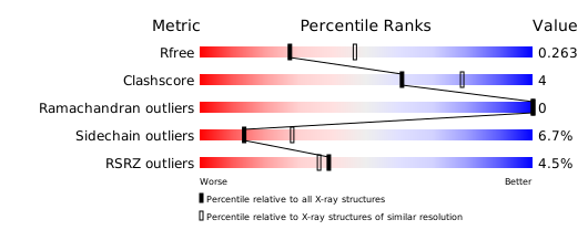
Structure validation
Download full validation report


