






 8WWX
8WWX
Ube1L acts akin to a mitt, that mediates UbcH8 binding and orchestrates "E1-E2" interaction
Summary for 8WWX
| Entry DOI | 10.2210/pdb8wwx/pdb |
| NMR Information | BMRB: 53398 |
| Descriptor | Ubiquitin-like modifier-activating enzyme 7 (1 entity in total) |
| Functional Keywords | ubiquitin fold domain, ube1l, uba7, ufd, beta-grasp domain, ubch8, ligase |
| Biological source | Homo sapiens (human) |
| Total number of polymer chains | 1 |
| Total formula weight | 10261.58 |
| Authors | Dag, C.,Elgin, E.S.,Lee, W.,Ziarek, J.J. (deposition date: 2023-10-27, release date: 2023-11-08, Last modification date: 2026-07-29) |
| Primary citation | Dag, C.,Lambert, M.,Kazar, A.E.,Kahraman, K.,Gocenler, O.,Lee, W.,Tozkoparan Ceylan, C.D.,Lohr, F.,Shim, J.G.,Haas, A.L.,Dotsch, V.,Ziarek, J.,Elgin, E.S. Dynamic Hotspots in the Uba7 Ubiquitin-Fold Domain Direct UbcH8 Recognition. Biochemistry, 65:678-692, 2026 Cited by PubMed Abstract: ISGylation is a ubiquitin-like post-translational modification that plays a central role in innate immune signaling. Conjugation of interferon-stimulated gene 15 (ISG15) to target proteins is initiated by the E1 enzyme Uba7, transferred to the E2 enzyme UbcH8, and completed by an E3 ligase. Specificity in this cascade is mediated by the ubiquitin-fold domain (UFD) of Uba7, yet the structural and mechanistic basis of E1-E2 recognition remains poorly defined. Here, we present the solution NMR structure and functional characterization of a human Uba7-UFD. NMR chemical shift perturbation experiments combined with site-directed mutagenesis delineate the UbcH8 interaction surface and identify residues critical for E1-E2 binding. The Uba7-UFD adopts a conserved ubiquitin-fold architecture but exhibits conformational flexibility in the unbound state. N relaxation measurements show a globally well-folded domain with localized ps-ns time scale dynamics within the β2/β4 E2 binding surface and the acidic loop spanning residues 996-1008. Upon UbcH8 binding, relaxation parameters shift toward those expected for a larger effective molecular size, accompanied by an increased residue-specific heterogeneity at the interface, consistent with binding-coupled changes in local mobility. Mutational analysis identifies C996 as being essential for UFD structural integrity and binding competence. Moreover, targeted alterations in the length and flexibility of the adjacent acidic loop strongly impair UbcH8 binding, demonstrating that the loop architecture is a critical determinant of efficient E2 recruitment. Together, these results provide a structural and dynamic framework for understanding E2 enzyme selection in the ISGylation pathway and highlight the role of UFD conformational dynamics in the E1-E2 complex formation. PubMed: 41773046DOI: 10.1021/acs.biochem.5c00807 PDB entries with the same primary citation |
| Experimental method | SOLUTION NMR |
Structure validation
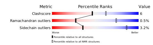
Download full validation report


