






 8V53
8V53
CryoEM structure of Ku homodimer in complex with hairpin DNA
Summary for 8V53
| Entry DOI | 10.2210/pdb8v53/pdb |
| EMDB information | 42978 |
| Descriptor | Non-homologous end joining protein Ku, DNA (21-MER), DNA (34-MER) (3 entities in total) |
| Functional Keywords | nhej, dna repair, mycobacterium tuberculosis, dna synapsis, dna binding protein |
| Biological source | Mycobacterium tuberculosis H37Rv More |
| Total number of polymer chains | 4 |
| Total formula weight | 84051.67 |
| Authors | Baral, J.,Rouiller, I.,Das, A.K. (deposition date: 2023-11-30, release date: 2025-12-10, Last modification date: 2026-04-08) |
| Primary citation | Baral, J.,Ang, C.S.,McMillan, P.J.,Shobhana, K.,Saini, A.,Hinde, E.,Das, A.K.,Rouiller, I. Bringing the ends together: cryo-EM structures of mycobacterial Ku in complex with DNA define its role in NHEJ synapsis. Nucleic Acids Res., 54:-, 2026 Cited by PubMed Abstract: Non-homologous end joining (NHEJ) is the sole pathway for repairing double-strand breaks in Mycobacterium tuberculosis during dormancy, relying on mycobacterial Ku (mKu) and ligase D, with mKu as the rate-limiting factor. Despite its essential role, the lack of structural information on prokaryotic Ku has hindered understanding of the molecular mechanisms underlying bacterial two-component NHEJ machinery. Here, we present the first cryo-electron microscopy (cryo-EM) structures of mKu in DNA-bound and higher-order supercomplex forms, revealing a Ku-mediated DNA synapsis mechanism unique to prokaryotes. Integrating cryo-EM with hydrogen-deuterium exchange mass spectrometry, we define key mKu-mKu dimerization, DNA-binding, and synapsis interactions essential for efficient NHEJ, bridging structure with function. Structure-guided in silico mutagenesis, coupled with electrophoretic mobility shift assays, identifies residues essential for DNA binding and synaptic assembly, which are crucial for NHEJ. Förster resonance energy transfer confirms DNA-dependent mKu oligomerization in solution, while live-cell imaging captures its spatiotemporal dynamics during double-stranded DNA break repair. These findings provide fundamental insights into the architecture and function of prokaryotic NHEJ, positioning mKu as a potential therapeutic target against tuberculosis and offering a framework for understanding DNA repair across bacterial species. PubMed: 41521670DOI: 10.1093/nar/gkaf1418 PDB entries with the same primary citation |
| Experimental method | ELECTRON MICROSCOPY (4.1 Å) |
Structure validation
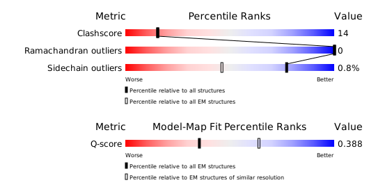
Download full validation report


