






 8TM8
8TM8
Monomer structure of monellin loop1 mutant (YENKG)
Summary for 8TM8
| Entry DOI | 10.2210/pdb8tm8/pdb |
| Related | 7D75 |
| Descriptor | Monellin chain B,Monellin chain A, SODIUM ION (3 entities in total) |
| Functional Keywords | single-chain monellin, sweet protein, domain swapping, plant protein |
| Biological source | Dioscoreophyllum cumminsii (serendipity berry) More |
| Total number of polymer chains | 2 |
| Total formula weight | 21418.47 |
| Authors | Manjula, R.,Pavithra, G.C.,Ramaswamy, S.,Gosavi, S. (deposition date: 2023-07-28, release date: 2024-12-18, Last modification date: 2026-07-22) |
| Primary citation | Manjula, R.,Chockalingam, N.,Subramanian, R.,Gosavi, S. On proline isomerization and 3D-domain-swapping. Biochem.Biophys.Res.Commun., 821:153872-153872, 2026 Cited by PubMed Abstract: 3D-domain-swapping is the exchange of identical "domains" between two protein monomers, which leads to homodimerization. This domain exchange can be facilitated by a single hinge-loop extending out. Hinge-loop prolines have been associated with domain-swapping, but their role remains unclear. Previously, we had engineered three domain-swapping variants of the monomeric monellin by replacing the wild-type hinge-loop sequence (L1:YENEGFREIKG) with QEVKG, YEIKG or QVVAG. The central residues of these sequences are hydrophobic (V/I), and lie at the apex of a tight solvent-exposed hinge-loop (L1Δ6) connecting two β-strands (β2-β3). The hydrophobic residue-solvent interaction likely impedes the closure of L1Δ6 and the formation of intra-chain β2-β3 contacts. This promotes domain-swapping. Here, we replaced the apex residue with the borderline-hydrophobic proline and found that it reduced domain-swapping in all three variants, with significant domain-swapping observed only in the most hydrophobic QVPAG construct. We then structurally characterized three monomeric (QEPKG, YEPKG, and QVPAG) and one domain-swapped dimeric (QVPAG) variants of monellin using X-ray crystallography. Interestingly, we find that the dimer has a trans-proline isomer, whereas all three monomers have a cis-proline. Thus, introducing proline into a solvent-exposed tight β-turn may be a robust method for designing cis-proline. Conversely, mutating such a naturally-occurring cis-proline to a hydrophobic amino acid may induce domain-swapping. The trans-proline in a hydrophobic hinge-loop (e.g. QVPAG), can provide rigidity to the domain-swapped dimer enabling the precise design of domain-swapping-driven protein assemblies. Overall, our mutational-design strategy is a step towards clarifying the role of prolines in 3D-domain-swapping and the rational design of proline isomerization. PubMed: 42102672DOI: 10.1016/j.bbrc.2026.153872 PDB entries with the same primary citation |
| Experimental method | X-RAY DIFFRACTION (2.26 Å) |
Structure validation
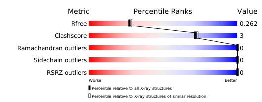
Download full validation report


