






 22MJ
22MJ
Crystal structure of human INPP5K with an allosteric inhibitor reveals the structural basis for species specific potency
This is a non-PDB format compatible entry.
Summary for 22MJ
| Entry DOI | 10.2210/pdb22mj/pdb |
| Descriptor | Inositol polyphosphate 5-phosphatase K, 1,2-ETHANEDIOL, MALONATE ION, ... (5 entities in total) |
| Functional Keywords | allosteric inihibitor complex, transferase |
| Biological source | Homo sapiens (human) |
| Total number of polymer chains | 1 |
| Total formula weight | 37944.39 |
| Authors | Nomura, A.,Yamaguchi, K.,Adachi, T. (deposition date: 2026-01-16, release date: 2026-03-11, Last modification date: 2026-04-15) |
| Primary citation | Nomura, A.,Yamaguchi, K.,Kawano, M.,Hanada, K.,Nishihata, J.,Noguchi, M.,Adachi, T. Crystal structure of human INPP5K with an allosteric inhibitor reveals the structural basis for species specific potency. Sci Rep, 16:-, 2026 Cited by PubMed Abstract: Inositol polyphosphate 5-phosphatase K (INPP5K) is a phosphatidylinositol (3,4,5)-trisphosphate phosphatase that increases glucose uptake and regulates myogenesis in the skeletal muscle. To understand the mechanism of its species-specific inhibition, we determined the 1.9-Å resolution crystal structure of human INPP5K in complex with a selective inhibitor, CPD-1 (IC = 2.9 µM). The structure reveals that CPD-1 binds to a novel allosteric pocket, inducing a large conformational change in α-helix 3 that alters the active site and prevents substrate binding. This finding explains its unique, noncompetitive inhibitory mechanism. Crucially, while the inhibitor-binding residues are conserved, the key residue governing the allosteric transition is not conserved in mouse and rat INPP5K, which correlates with their insensitivity to CPD-1 (IC > 100 µM). Based on these structural insights, we identified the hamster as a pharmacologically relevant preclinical model (IC = 8.2 µM). These findings provide a structural basis for the rational design of next-generation INPP5K inhibitors and establish a suitable animal model for their evaluation. PubMed: 41748720DOI: 10.1038/s41598-026-40748-4 PDB entries with the same primary citation |
| Experimental method | X-RAY DIFFRACTION (1.9 Å) |
Structure validation
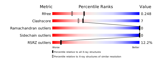
Download full validation report


