9JDQ
 
 | |
9JDT
 | |
9JD2
 | |
9DE2
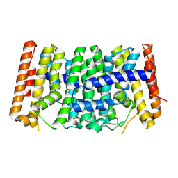
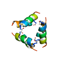
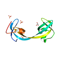
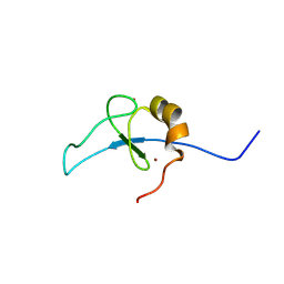 | | ETO2 MYND bound to MPPL peptide from GATAD2A | | 分子名称: | GATAD2A-MPPL motif and ETO2 NHR4 domain fusion protein, ZINC ION | | 著者 | Williams, D.C. | | 登録日 | 2024-08-28 | | 公開日 | 2024-09-18 | | 実験手法 | SOLUTION NMR | | 主引用文献 | The role of multivalency in the association of the eight twenty-one protein 2 (ETO2) with the nucleosome remodeling and deacetylase (NuRD) complex
To Be Published
|
|
9DDY
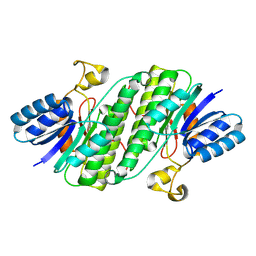
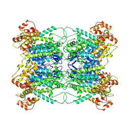
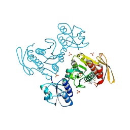
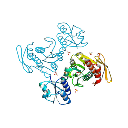
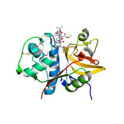
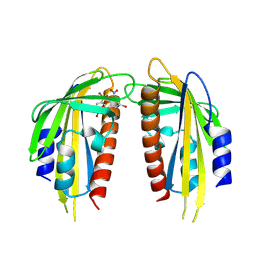
 | | The Crystal Structure of Geranyltranstransferase from Streptococcus pneumoniae TIGR4 | | 分子名称: | GLYCEROL, Geranyltranstransferase | | 著者 | Kim, Y, Tan, A, Endres, M, Tan, K, Joachimik, A, Center for Structural Biology of Infectious Diseases (CSBID) | | 登録日 | 2024-08-28 | | 公開日 | 2024-09-18 | | 実験手法 | X-RAY DIFFRACTION (2.32 Å) | | 主引用文献 | The Crystal Structure of Geranyltranstransferase from Streptococcus pneumoniae TIGR4
To Be Published
|
|
9JBQ
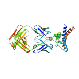 | | Structure of the complex between h1F3 Fab and PcrV fragment | | 分子名称: | Heavy chain, PcrV, light chain | | 著者 | Numata, S, Hara, T, Izawa, M, Okuno, Y, Sato, Y, Yamane, S, Maki, H, Sato, T, Yamano, Y. | | 登録日 | 2024-08-27 | | 公開日 | 2024-09-11 | | 実験手法 | X-RAY DIFFRACTION (2 Å) | | 主引用文献 | Novel humanized anti-PcrV monoclonal antibody, COT-143, protects mice from lethal Pseudomonas aeruginosa infection via inhibition of toxin translocation by the type III secretion system
To Be Published
|
|
9GLE
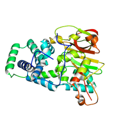
 | | Jumonji domain-containing protein 2A with crystallization epitope mutations A91T:T93S | | 分子名称: | 1,2-ETHANEDIOL, Lysine-specific demethylase 4A, NICKEL (II) ION, ... | | 著者 | Fairhead, M, Strain-Damerell, C, Ye, M, Mackinnon, S.R, Pinkas, D, MacLean, E.M, Koekemoer, L, Damerell, D, Krojer, T, Arrowsmith, C.H, Edwards, A, Bountra, C, Yue, W, Burgess-Brown, N, Marsden, B, von Delft, F, Structural Genomics Consortium (SGC) | | 登録日 | 2024-08-27 | | 公開日 | 2024-09-18 | | 実験手法 | X-RAY DIFFRACTION (1.88 Å) | | 主引用文献 | A fast, parallel method for efficiently exploring crystallization behaviour of large numbers of protein variants
To be published
|
|
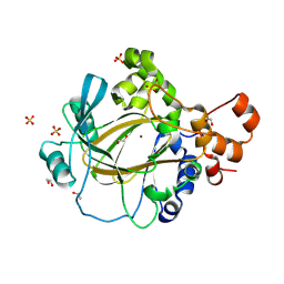
9DCG
 | | Crystal Structure of the Thiol:Disulfide Interchange Protein DsbC from Vibrio cholerae | | 分子名称: | 1,2-ETHANEDIOL, CHLORIDE ION, FORMIC ACID, ... | | 著者 | Kim, Y, Maltseva, N, Shatsman, S, Joachimiak, A, Center for Structural Biology of Infectious Diseases (CSBID) | | 登録日 | 2024-08-26 | | 公開日 | 2024-09-04 | | 実験手法 | X-RAY DIFFRACTION (2.09 Å) | | 主引用文献 | Crystal Structure of the Thiol:Disulfide Interchange Protein DsbC from Vibrio cholerae
To Be Published
|
|
9JA1
 | |
9JA0
 | |
9J9K
 | |
9J9A
 | | Staphylococcus aureus exfoliative toxin D | | 分子名称: | SULFATE ION, Serine protease, beta-D-glucopyranose | | 著者 | Tonozuka, T. | | 登録日 | 2024-08-22 | | 公開日 | 2024-09-11 | | 最終更新日 | 2024-09-18 | | 実験手法 | X-RAY DIFFRACTION (1.75 Å) | | 主引用文献 | Crystal structure of exfoliative toxin D from Staphylococcus aureus
Biochem.Biophys.Res.Commun., 2024
|
|
9J9C
 | |
9D9K
 | |
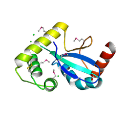
9J8F
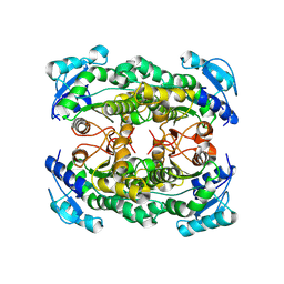
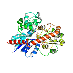
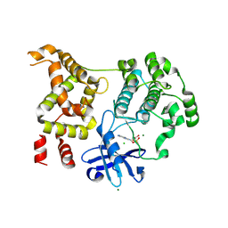
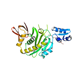 | | Structural insights into BirA from Haemophilus influenzae, a bifunctional protein as a biotin protein ligase and a transcriptional repressor | | 分子名称: | 3,6,9,12,15,18-HEXAOXAICOSANE-1,20-DIOL, Bifunctional ligase/repressor BirA, PENTAETHYLENE GLYCOL | | 著者 | Lee, J.Y, Jeong, K.H, Son, S.B, Ko, J.H. | | 登録日 | 2024-08-21 | | 公開日 | 2024-09-11 | | 実験手法 | X-RAY DIFFRACTION (2.65 Å) | | 主引用文献 | Structural insights into BirA from Haemophilus influenzae, a bifunctional protein as a biotin protein ligase and a transcriptional repressor.
Biochem.Biophys.Res.Commun., 733, 2024
|
|
9D9M
 | |
9J8E
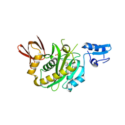 | | Structural insights into BirA from Haemophilus influenzae, a bifunctional protein as a biotin protein ligase and a transcriptional repressor | | 分子名称: | BIOTINYL-5-AMP, Bifunctional ligase/repressor BirA | | 著者 | Lee, J.Y, Jeong, K.H, Son, S.B, Ko, J.H. | | 登録日 | 2024-08-21 | | 公開日 | 2024-09-11 | | 実験手法 | X-RAY DIFFRACTION (2.65 Å) | | 主引用文献 | Structural insights into BirA from Haemophilus influenzae, a bifunctional protein as a biotin protein ligase and a transcriptional repressor.
Biochem.Biophys.Res.Commun., 733, 2024
|
|
9GJ2
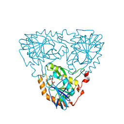
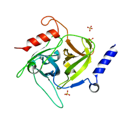
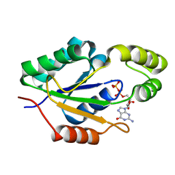
 | | Crystal structure of human cathepsin S produced in insect cells in complex with ketoamide 13b | | 分子名称: | 1,2-ETHANEDIOL, CITRIC ACID, Cathepsin S, ... | | 著者 | Falke, S, Karnicar, K, Usenik, A, Lindic, N, Sekirnik, A, Reinke, P.Y.A, Guenther, S, Turk, D, Meents, A. | | 登録日 | 2024-08-20 | | 公開日 | 2024-09-04 | | 実験手法 | X-RAY DIFFRACTION (1.15 Å) | | 主引用文献 | Crystal structure of human cathepsin S produced in insect cells in complex with ketoamide 13b
To Be Published
|
|
9D8S
 | |
9J7T
 | |
9D8A
 | |
9GII
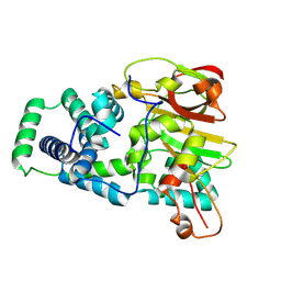
 | | Jumonji domain-containing protein 2A with crystallization epitope mutation R913A | | 分子名称: | GLYCEROL, Lysine-specific demethylase 4A, SULFATE ION | | 著者 | Fairhead, M, Strain-Damerell, C, Ye, M, Mackinnon, S.R, Pinkas, D, MacLean, E.M, Koekemoer, L, Damerell, D, Krojer, T, Arrowsmith, C.H, Edwards, A, Bountra, C, Yue, W, Burgess-Brown, N, Marsden, B, von Delft, F, Structural Genomics Consortium (SGC) | | 登録日 | 2024-08-19 | | 公開日 | 2024-09-04 | | 実験手法 | X-RAY DIFFRACTION (1.7 Å) | | 主引用文献 | A fast, parallel method for efficiently exploring crystallization behaviour of large numbers of protein variants
To be published
|
|
9J6I
 | |
9D6J
 | | Nitrile hydratase BR52K mutant | | 分子名称: | Cobalt-containing nitrile hydratase subunit alpha, Cobalt-containing nitrile hydratase subunit beta | | 著者 | Miller, C.G, Holz, R.C, Liu, D, Kaley, N. | | 登録日 | 2024-08-15 | | 公開日 | 2024-08-28 | | 実験手法 | X-RAY DIFFRACTION (1.47 Å) | | 主引用文献 | Role of second-sphere arginine residues in metal binding and metallocentre assembly in nitrile hydratases.
J Inorg Biochem, 256, 2024
|
|
9D6M
 | | Nitrile hydratase BR157K mutant | | 分子名称: | Cobalt-containing nitrile hydratase subunit alpha, Cobalt-containing nitrile hydratase subunit beta | | 著者 | Miller, C.G, Holz, R.C, Liu, D, Kaley, N. | | 登録日 | 2024-08-15 | | 公開日 | 2024-08-28 | | 実験手法 | X-RAY DIFFRACTION (1.56 Å) | | 主引用文献 | Role of second-sphere arginine residues in metal binding and metallocentre assembly in nitrile hydratases.
J Inorg Biochem, 256, 2024
|
|


