Loading
PDBj
MenuPDBj@FacebookPDBj@TwitterPDBj@YouTubewwPDB FoundationwwPDB
RCSB PDBPDBeBMRBAdv. SearchSearch help

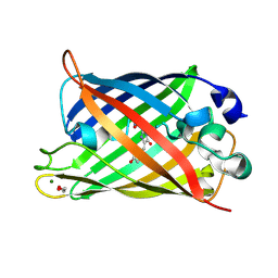
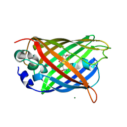
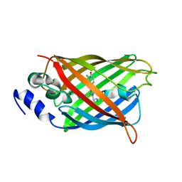
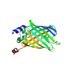
6PFU
DownloadVisualize
BU of 6pfu by Molmil
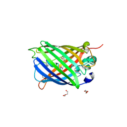
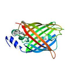
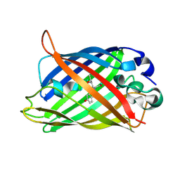
rsEGFP2 with a chlorinated chromophore in the non-fluorescent off-state in a contracted unit cell
Descriptor: Green fluorescent protein, SULFATE ION
Authors:Chang, J, Romei, M.G, Boxer, S.G.
Deposit date:2019-06-22
Release date:2019-08-07
Last modified:2023-11-15
Method:X-RAY DIFFRACTION (1.619 Å)
Cite:Structural Evidence of Photoisomerization Pathways in Fluorescent Proteins.
J.Am.Chem.Soc., 141, 2019
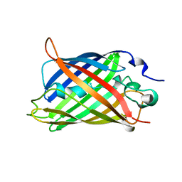
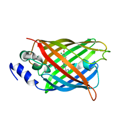
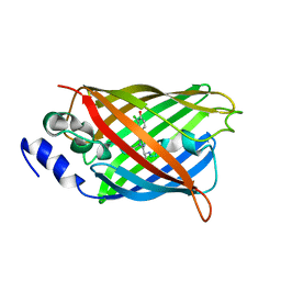
6PFR
DownloadVisualize
BU of 6pfr by Molmil
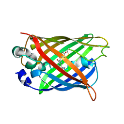
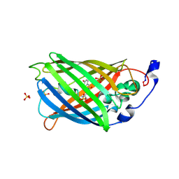
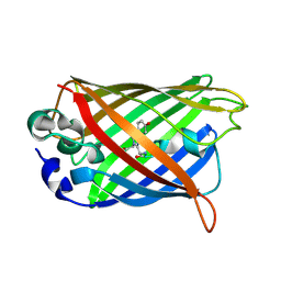
rsEGFP2 with a chlorinated chromophore in the fluorescent on-state
Descriptor: Green fluorescent protein
Authors:Chang, J, Romei, M.G, Boxer, S.G.
Deposit date:2019-06-22
Release date:2019-08-07
Last modified:2023-11-15
Method:X-RAY DIFFRACTION (1.51 Å)
Cite:Structural Evidence of Photoisomerization Pathways in Fluorescent Proteins.
J.Am.Chem.Soc., 141, 2019
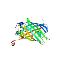
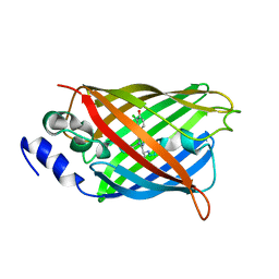
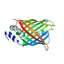
2HRS
DownloadVisualize
BU of 2hrs by Molmil
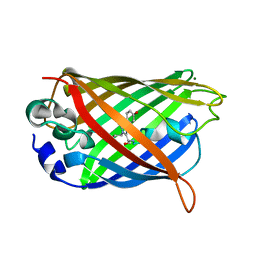
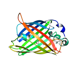
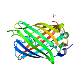
Crystal structure of L42H V224H design intermediate for GFP metal ion reporter
Descriptor: 1,2-ETHANEDIOL, Green fluorescent protein, MAGNESIUM ION
Authors:Barondeau, D.P, Tubbs, J.L, Tainer, J.A, Getzoff, E.D.
Deposit date:2006-07-20
Release date:2008-04-08
Last modified:2023-11-15
Method:X-RAY DIFFRACTION (1.4 Å)
Cite:Iterative Structure-Based Design of a Green Fluorescent Protein Metal Ion Reporter
To be Published
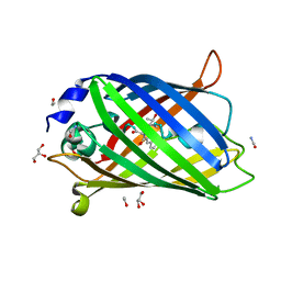
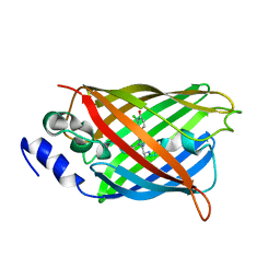
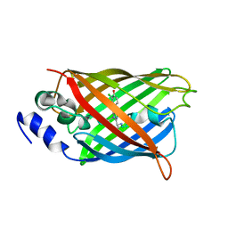
1GFL
DownloadVisualize
BU of 1gfl by Molmil
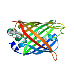
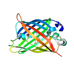
STRUCTURE OF GREEN FLUORESCENT PROTEIN
Descriptor: GREEN FLUORESCENT PROTEIN
Authors:Yang, F, Moss, L.G, Phillips Jr, G.N.
Deposit date:1996-08-23
Release date:1997-01-11
Last modified:2024-04-24
Method:X-RAY DIFFRACTION (1.9 Å)
Cite:The molecular structure of green fluorescent protein.
Nat.Biotechnol., 14, 1996
7PCZ
DownloadVisualize
BU of 7pcz by Molmil
Functional and structural characterization of redox sensitive superfolder green fluorescent protein and variants
Descriptor: ETHANOL, GLYCEROL, Green fluorescent protein
Authors:Fritz-Wolf, K, Heimsch, K.C, Schuh, A.K, Becker, K.
Deposit date:2021-08-04
Release date:2022-02-16
Last modified:2024-01-31
Method:X-RAY DIFFRACTION (1.35 Å)
Cite:Structure and Function of Redox-Sensitive Superfolder Green Fluorescent Protein Variant.
Antioxid.Redox Signal., 37, 2022
7PCA
DownloadVisualize
BU of 7pca by Molmil
Functional and structural characterization of redox sensitive superfolder green fluorescent protein and variants
Descriptor: ETHANOL, FORMAMIDE, GLYCEROL, ...
Authors:Fritz-Wolf, K, Heimsch, K.C, Schuh, A.K, Becker, K.
Deposit date:2021-08-03
Release date:2022-02-16
Last modified:2024-01-31
Method:X-RAY DIFFRACTION (1.05 Å)
Cite:Structure and Function of Redox-Sensitive Superfolder Green Fluorescent Protein Variant.
Antioxid.Redox Signal., 37, 2022
7PD0
DownloadVisualize
BU of 7pd0 by Molmil
Functional and structural characterization of redox sensitive superfolder green fluorescent protein and variants
Descriptor: DI(HYDROXYETHYL)ETHER, GLYCEROL, Green fluorescent protein, ...
Authors:Fritz-Wolf, K, Heimsch, K.C, Schuh, A.K, Becker, K.
Deposit date:2021-08-04
Release date:2022-02-16
Last modified:2024-01-31
Method:X-RAY DIFFRACTION (2 Å)
Cite:Structure and Function of Redox-Sensitive Superfolder Green Fluorescent Protein Variant.
Antioxid.Redox Signal., 37, 2022
3CD1
DownloadVisualize
BU of 3cd1 by Molmil
Development of a family of redox-sensitive green fluorescent protein indicators for use in relatively oxidizing subcellular environments
Descriptor: Green fluorescent protein
Authors:Lohman, J.R.
Deposit date:2008-02-26
Release date:2008-08-19
Last modified:2021-10-20
Method:X-RAY DIFFRACTION (1.312 Å)
Cite:Development of a family of redox-sensitive green fluorescent protein indicators for use in relatively oxidizing subcellular environments
Biochemistry, 47, 2008
3CBE
DownloadVisualize
BU of 3cbe by Molmil
Development of a family of redox-sensitive green fluorescent protein indicators for use in relatively oxidizing subcellular environments
Descriptor: Green fluorescent protein, MAGNESIUM ION
Authors:Lohman, J.R.
Deposit date:2008-02-21
Release date:2008-08-19
Last modified:2021-10-20
Method:X-RAY DIFFRACTION (1.488 Å)
Cite:Development of a family of redox-sensitive green fluorescent protein indicators for use in relatively oxidizing subcellular environments
Biochemistry, 47, 2008
3CD9
DownloadVisualize
BU of 3cd9 by Molmil
Development of a family of redox-sensitive green fluorescent protein indicators for use in relatively oxidizing subcellular environments
Descriptor: Green fluorescent protein
Authors:Lohman, J.R.
Deposit date:2008-02-26
Release date:2008-08-19
Last modified:2021-10-20
Method:X-RAY DIFFRACTION (1.502 Å)
Cite:Development of a family of redox-sensitive green fluorescent protein indicators for use in relatively oxidizing subcellular environments
Biochemistry, 47, 2008
3CB9
DownloadVisualize
BU of 3cb9 by Molmil
Development of a family of redox-sensitive green fluorescent protein indicators for use in relatively oxidizing subcellular environments
Descriptor: Green fluorescent protein, MAGNESIUM ION
Authors:Lohman, J.R.
Deposit date:2008-02-21
Release date:2008-08-19
Last modified:2023-11-15
Method:X-RAY DIFFRACTION (1.31 Å)
Cite:Development of a family of redox-sensitive green fluorescent protein indicators for use in relatively oxidizing subcellular environments
Biochemistry, 47, 2008
8A6O
DownloadVisualize
BU of 8a6o by Molmil
Room temperature rsEGFP2 with a chlorinated chromophore 600 fs after Photoexcitation
Descriptor: Green fluorescent protein
Authors:Fadini, A, van Thor, J.
Deposit date:2022-06-18
Release date:2023-07-19
Last modified:2024-02-07
Method:X-RAY DIFFRACTION (1.63 Å)
Cite:Serial Femtosecond Crystallography Reveals that Photoactivation in a Fluorescent Protein Proceeds via the Hula Twist Mechanism.
J.Am.Chem.Soc., 145, 2023
8A6S
DownloadVisualize
BU of 8a6s by Molmil
Room temperature rsEGFP2 with a chlorinated chromophore 1 microsecond after Photoexcitation
Descriptor: Green fluorescent protein
Authors:Fadini, A, van Thor, J.
Deposit date:2022-06-19
Release date:2023-07-19
Last modified:2024-02-07
Method:X-RAY DIFFRACTION (1.63 Å)
Cite:Serial Femtosecond Crystallography Reveals that Photoactivation in a Fluorescent Protein Proceeds via the Hula Twist Mechanism.
J.Am.Chem.Soc., 145, 2023
8A6N
DownloadVisualize
BU of 8a6n by Molmil
Room temperature rsEGFP2 with a chlorinated chromophore 300 fs after Photoexcitation
Descriptor: Green fluorescent protein
Authors:Fadini, A, van Thor, J.
Deposit date:2022-06-18
Release date:2023-07-19
Last modified:2024-02-07
Method:X-RAY DIFFRACTION (1.63 Å)
Cite:Serial Femtosecond Crystallography Reveals that Photoactivation in a Fluorescent Protein Proceeds via the Hula Twist Mechanism.
J.Am.Chem.Soc., 145, 2023
8A6R
DownloadVisualize
BU of 8a6r by Molmil
Room temperature rsEGFP2 with a chlorinated chromophore 100 ps after Photoexcitation
Descriptor: Green fluorescent protein
Authors:Fadini, A, van Thor, J.
Deposit date:2022-06-18
Release date:2023-07-19
Last modified:2024-02-07
Method:X-RAY DIFFRACTION (1.63 Å)
Cite:Serial Femtosecond Crystallography Reveals that Photoactivation in a Fluorescent Protein Proceeds via the Hula Twist Mechanism.
J.Am.Chem.Soc., 145, 2023
8A6G
DownloadVisualize
BU of 8a6g by Molmil
Room temperature rsEGFP2 with a chlorinated chromophore in the non-fluorescent OFF-state
Descriptor: Green fluorescent protein
Authors:Fadini, A, van Thor, J.
Deposit date:2022-06-17
Release date:2023-07-19
Last modified:2024-02-07
Method:X-RAY DIFFRACTION (1.63 Å)
Cite:Serial Femtosecond Crystallography Reveals that Photoactivation in a Fluorescent Protein Proceeds via the Hula Twist Mechanism.
J.Am.Chem.Soc., 145, 2023
8A6P
DownloadVisualize
BU of 8a6p by Molmil
Room temperature rsEGFP2 with a chlorinated chromophore 900 fs after photoexcitation
Descriptor: Green fluorescent protein
Authors:Fadini, A, van Thor, J.
Deposit date:2022-06-18
Release date:2023-07-19
Last modified:2024-02-07
Method:X-RAY DIFFRACTION (1.63 Å)
Cite:Serial Femtosecond Crystallography Reveals that Photoactivation in a Fluorescent Protein Proceeds via the Hula Twist Mechanism.
J.Am.Chem.Soc., 145, 2023
8A6Q
DownloadVisualize
BU of 8a6q by Molmil
Room temperature rsEGFP2 with a chlorinated chromophore 5 ps after photoexcitation
Descriptor: Green fluorescent protein
Authors:Fadini, A, van Thor, J.
Deposit date:2022-06-18
Release date:2023-07-19
Last modified:2024-02-07
Method:X-RAY DIFFRACTION (1.63 Å)
Cite:Serial Femtosecond Crystallography Reveals that Photoactivation in a Fluorescent Protein Proceeds via the Hula Twist Mechanism.
J.Am.Chem.Soc., 145, 2023
8A7V
DownloadVisualize
BU of 8a7v by Molmil
Room temperature rsEGFP2 in its OFF-state obtained with SFX
Descriptor: Green fluorescent protein
Authors:Fadini, A, van Thor, J.
Deposit date:2022-06-21
Release date:2023-07-19
Last modified:2024-02-07
Method:X-RAY DIFFRACTION (1.46 Å)
Cite:Serial Femtosecond Crystallography Reveals that Photoactivation in a Fluorescent Protein Proceeds via the Hula Twist Mechanism.
J.Am.Chem.Soc., 145, 2023
1KP5
DownloadVisualize
BU of 1kp5 by Molmil
Cyclic Green Fluorescent Protein
Descriptor: Green Fluorescent Protein, SULFATE ION
Authors:Hofmann, A, Iwai, H, Plueckthun, A, Wlodawer, A.
Deposit date:2001-12-28
Release date:2002-08-28
Last modified:2023-11-15
Method:X-RAY DIFFRACTION (2.6 Å)
Cite:Structure of cyclized green fluorescent protein.
Acta Crystallogr.,Sect.D, 58, 2002
1F09
DownloadVisualize
BU of 1f09 by Molmil
CRYSTAL STRUCTURE OF THE GREEN FLUORESCENT PROTEIN (GFP) VARIANT YFP-H148Q WITH TWO BOUND IODIDES
Descriptor: GREEN FLUORESCENT PROTEIN, IODIDE ION
Authors:Wachter, R.M, Yarbrough, D, Kallio, K, Remington, S.J.
Deposit date:2000-05-15
Release date:2000-11-17
Last modified:2021-11-03
Method:X-RAY DIFFRACTION (2.14 Å)
Cite:Crystallographic and energetic analysis of binding of selected anions to the yellow variants of green fluorescent protein.
J.Mol.Biol., 301, 2000
1F0B
DownloadVisualize
BU of 1f0b by Molmil
CRYSTAL STRUCTURE OF THE GREEN FLUORESCENT PROTEIN (GFP) VARIANT YFP-H148Q
Descriptor: GREEN FLUORESCENT PROTEIN
Authors:Wachter, R.M, Yarbrough, D, Kallio, K, Remington, S.J.
Deposit date:2000-05-15
Release date:2000-11-17
Last modified:2021-11-03
Method:X-RAY DIFFRACTION (2.1 Å)
Cite:Crystallographic and energetic analysis of binding of selected anions to the yellow variants of green fluorescent protein.
J.Mol.Biol., 301, 2000
7YDQ
DownloadVisualize
BU of 7ydq by Molmil
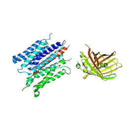
Structure of PfNT1(Y190A)-GFP in complex with GSK4
Descriptor: 5-methyl-N-[2-(2-oxidanylideneazepan-1-yl)ethyl]-2-phenyl-1,3-oxazole-4-carboxamide, Nucleoside transporter 1,Green fluorescent protein
Authors:Wang, C, Yu, L.Y, Li, J.L, Ren, R.B, Deng, D.
Deposit date:2022-07-04
Release date:2023-04-26
Last modified:2024-07-03
Method:ELECTRON MICROSCOPY (4.04 Å)
Cite:Structural basis of the substrate recognition and inhibition mechanism of Plasmodium falciparum nucleoside transporter PfENT1.
Nat Commun, 14, 2023
2DUF
DownloadVisualize
BU of 2duf by Molmil
crystal structure of a green fluorescent protein variant S65T/H148D at pH 5.6
Descriptor: Green fluorescent protein
Authors:Shu, X, Remington, S.J.
Deposit date:2006-07-23
Release date:2007-07-10
Last modified:2024-07-10
Method:X-RAY DIFFRACTION (1.5 Å)
Cite:Ultrafast excited-state dynamics in the green fluorescent protein variant S65T/H148D. 1. Mutagenesis and structural studies.
Biochemistry, 46, 2007
2DUI
DownloadVisualize
BU of 2dui by Molmil
crystal structure of a green fluorescent protein variant H148D at pH 9
Descriptor: Green fluorescent protein
Authors:Shu, X, Remington, S.J.
Deposit date:2006-07-23
Release date:2007-07-10
Last modified:2023-11-15
Method:X-RAY DIFFRACTION (1.36 Å)
Cite:Ultrafast excited-state dynamics in the green fluorescent protein variant S65T/H148D. 1. Mutagenesis and structural studies.
Biochemistry, 46, 2007

225399

건을2024-09-25부터공개중

PDB statisticsPDBj update infoContact PDBjnumon