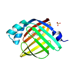
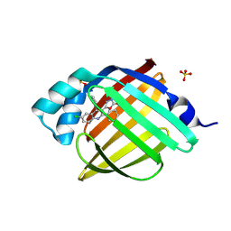
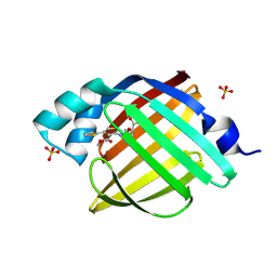
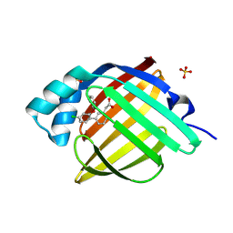
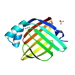
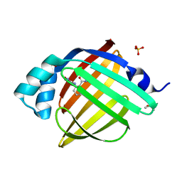
7G02
 
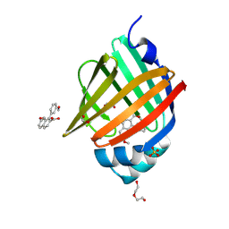 | | Crystal Structure of human FABP4 in complex with 7-methoxy-1-(3-methoxyphenyl)naphthalene-2-carboxylic acid, i.e. SMILES c1(c2c(ccc1C(=O)O)ccc(c2)OC)c1cc(ccc1)OC with IC50=0.045 microM | | Descriptor: | (1M)-7-methoxy-1-(3-methoxyphenyl)naphthalene-2-carboxylic acid, 1,2-ETHANEDIOL, FORMIC ACID, ... | | Authors: | Ehler, A, Benz, J, Obst, U, Guthrie-Robert, W, Rudolph, M.G. | | Deposit date: | 2023-04-27 | | Release date: | 2023-06-14 | | Last modified: | 2025-08-13 | | Method: | X-RAY DIFFRACTION (1.03 Å) | | Cite: | A high-resolution data set of fatty acid-binding protein structures. III. Unexpectedly high occurrence of wrong ligands.
Acta Crystallogr D Struct Biol, 81, 2025
|
|
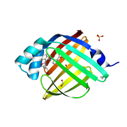
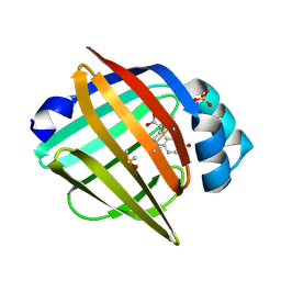
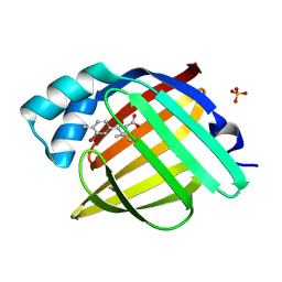
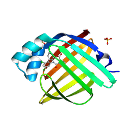
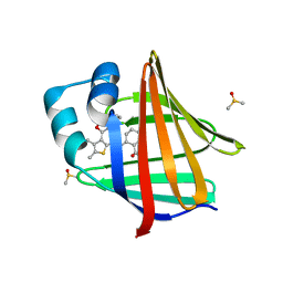
7G03
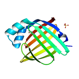 | | Crystal Structure of human FABP4 in complex with 6-fluoro-1,3-benzothiazol-2-amine | | Descriptor: | 6-fluoro-1,3-benzothiazol-2-amine, Fatty acid-binding protein, adipocyte, ... | | Authors: | Ehler, A, Benz, J, Obst, U, Winternitz, P, Rudolph, M.G. | | Deposit date: | 2023-04-27 | | Release date: | 2023-06-14 | | Last modified: | 2025-08-13 | | Method: | X-RAY DIFFRACTION (1.27 Å) | | Cite: | A high-resolution data set of fatty acid-binding protein structures. III. Unexpectedly high occurrence of wrong ligands.
Acta Crystallogr D Struct Biol, 81, 2025
|
|
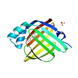
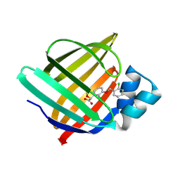
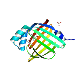
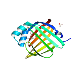
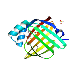
7G04
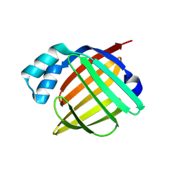 | | Crystal Structure of human FABP5 in complex with 7-(4-chlorophenyl)-1,2,3,4-tetrahydronaphthalene-1-carboxylic acid, i.e. SMILES c12c(ccc(c2)c2ccc(cc2)Cl)CCC[C@H]1C(=O)O with IC50=3.3 microM | | Descriptor: | (1R)-7-(4-chlorophenyl)-1,2,3,4-tetrahydronaphthalene-1-carboxylic acid, CHLORIDE ION, DIMETHYL SULFOXIDE, ... | | Authors: | Ehler, A, Benz, J, Obst, U, Canesso, R, Rudolph, M.G. | | Deposit date: | 2023-04-27 | | Release date: | 2023-06-14 | | Last modified: | 2025-08-13 | | Method: | X-RAY DIFFRACTION (1.4 Å) | | Cite: | A high-resolution data set of fatty acid-binding protein structures. III. Unexpectedly high occurrence of wrong ligands.
Acta Crystallogr D Struct Biol, 81, 2025
|
|
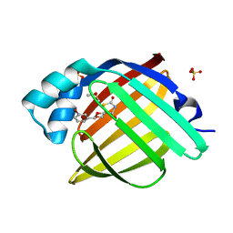
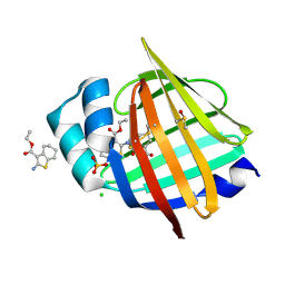
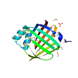
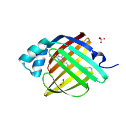
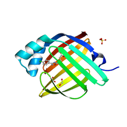
7G05
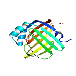 | | Crystal Structure of human FABP4 in complex with 2,6-dichloro-4-(1,1,1,3,3,3-hexafluoro-2-hydroxypropan-2-yl)phenol, i.e. SMILES C(C(F)(F)F)(C(F)(F)F)(c1cc(c(c(c1)Cl)O)Cl)O with IC50=12 microM | | Descriptor: | 2,6-dichloro-4-(1,1,1,3,3,3-hexafluoro-2-hydroxypropan-2-yl)phenol, DIMETHYL SULFOXIDE, Fatty acid-binding protein, ... | | Authors: | Ehler, A, Benz, J, Obst, U, Rudolph, M.G. | | Deposit date: | 2023-04-27 | | Release date: | 2023-06-14 | | Last modified: | 2025-08-13 | | Method: | X-RAY DIFFRACTION (0.95 Å) | | Cite: | A high-resolution data set of fatty acid-binding protein structures. III. Unexpectedly high occurrence of wrong ligands.
Acta Crystallogr D Struct Biol, 81, 2025
|
|
7G06
 | | Crystal Structure of human FABP4 in complex with 5-(chloromethyl)-2-phenyl-4H-[1,2,4]triazolo[1,5-a]pyrimidin-7-one, i.e. SMILES c1cccc(c1)C1=NN2C(=N1)NC(=CC2=O)CCl with IC50=28 microM | | Descriptor: | (8S)-5-(chloromethyl)-2-phenyl[1,2,4]triazolo[1,5-a]pyrimidin-7(4H)-one, Fatty acid-binding protein, adipocyte, ... | | Authors: | Ehler, A, Benz, J, Obst, U, Ceccarelli-Simona, M, Rudolph, M.G. | | Deposit date: | 2023-04-27 | | Release date: | 2023-06-14 | | Last modified: | 2025-08-13 | | Method: | X-RAY DIFFRACTION (1.26 Å) | | Cite: | A high-resolution data set of fatty acid-binding protein structures. III. Unexpectedly high occurrence of wrong ligands.
Acta Crystallogr D Struct Biol, 81, 2025
|
|
7G07
 | | Crystal Structure of human FABP4 in complex with 4-[[3-(3-cyclopropyl-1,2,4-oxadiazol-5-yl)-4,5,6,7-tetrahydro-1-benzothiophen-2-yl]carbamoyl]-3,6-dihydro-2H-pyran-5-carboxylic acid, i.e. SMILES S1C(=C(C2=NC(=NO2)C2CC2)C2=C1CCCC2)NC(=O)C1=C(COCC1)C(=O)O with IC50=0.0174352 microM | | Descriptor: | 4-{[(3M)-3-(3-cyclopropyl-1,2,4-oxadiazol-5-yl)-4,5,6,7-tetrahydro-1-benzothiophen-2-yl]carbamoyl}-5,6-dihydro-2H-pyran-3-carboxylic acid, DIMETHYL SULFOXIDE, FORMIC ACID, ... | | Authors: | Ehler, A, Benz, J, Obst, U, Richter, H, Rudolph, M.G. | | Deposit date: | 2023-04-27 | | Release date: | 2023-06-14 | | Last modified: | 2025-08-13 | | Method: | X-RAY DIFFRACTION (1.61 Å) | | Cite: | A high-resolution data set of fatty acid-binding protein structures. III. Unexpectedly high occurrence of wrong ligands.
Acta Crystallogr D Struct Biol, 81, 2025
|
|
7G08
 | | Crystal Structure of human FABP4 in complex with 2-(3-chloro-2-methylanilino)benzoic acid, i.e. SMILES c1(c(cccc1)C(=O)O)Nc1c(c(ccc1)Cl)C with IC50=0.482076 microM | | Descriptor: | 2-[(3-chloro-2-methylphenyl)amino]benzoic acid, Fatty acid-binding protein, adipocyte, ... | | Authors: | Ehler, A, Benz, J, Obst, U, Boehringer, M, Rudolph, M.G. | | Deposit date: | 2023-04-27 | | Release date: | 2023-06-14 | | Last modified: | 2025-08-13 | | Method: | X-RAY DIFFRACTION (0.99 Å) | | Cite: | A high-resolution data set of fatty acid-binding protein structures. III. Unexpectedly high occurrence of wrong ligands.
Acta Crystallogr D Struct Biol, 81, 2025
|
|
7G09
 | | Crystal Structure of human FABP4 in complex with 8-(3-bromophenyl)-7,9-dioxa-1-thia-3-azaspiro[4.5]decane-2,4-dione, i.e. SMILES [C@@]12(C(=O)NC(=O)S1)CO[C@@H](OC2)c1cc(ccc1)Br with IC50=2.3 microM | | Descriptor: | (5s,8s)-8-(3-bromophenyl)-7,9-dioxa-1-thia-3-azaspiro[4.5]decane-2,4-dione, DIMETHYL SULFOXIDE, FORMIC ACID, ... | | Authors: | Ehler, A, Benz, J, Obst, U, Kumagai, Y, Rudolph, M.G. | | Deposit date: | 2023-04-27 | | Release date: | 2023-06-14 | | Last modified: | 2025-08-13 | | Method: | X-RAY DIFFRACTION (1.05 Å) | | Cite: | A high-resolution data set of fatty acid-binding protein structures. III. Unexpectedly high occurrence of wrong ligands.
Acta Crystallogr D Struct Biol, 81, 2025
|
|
7G0A
 | | Crystal Structure of human FABP4 in complex with 5-(6-chloro-4-phenyl-2-piperidin-1-ylquinolin-3-yl)-3H-1,3,4-oxadiazol-2-one, i.e. SMILES c1(ccc2c(c1)c(c(c(n2)N1CCCCC1)C1=NNC(=O)O1)c1ccccc1)Cl with IC50=0.070 microM | | Descriptor: | (5M)-5-[6-chloro-4-phenyl-2-(piperidin-1-yl)quinolin-3-yl]-1,3,4-oxadiazol-2(3H)-one, Fatty acid-binding protein, adipocyte, ... | | Authors: | Ehler, A, Benz, J, Obst, U, Kuhne, H, Rudolph, M.G. | | Deposit date: | 2023-04-27 | | Release date: | 2023-06-14 | | Last modified: | 2025-08-13 | | Method: | X-RAY DIFFRACTION (1.12 Å) | | Cite: | A high-resolution data set of fatty acid-binding protein structures. III. Unexpectedly high occurrence of wrong ligands.
Acta Crystallogr D Struct Biol, 81, 2025
|
|
7G0B
 | | Crystal Structure of human FABP5 in complex with 7-bromo-1-methyl-5-phenyl-2,3,4,5-tetrahydro-1-benzazepine-4-carboxylic acid, i.e. SMILES [C@@H]1([C@@H](CCN(c2c1cc(cc2)Br)C)C(=O)O)c1ccccc1 with IC50=2.3 microM | | Descriptor: | (4S,5S)-7-bromo-1-methyl-5-phenyl-2,3,4,5-tetrahydro-1H-1-benzazepine-4-carboxylic acid, DIMETHYL SULFOXIDE, Fatty acid-binding protein 5, ... | | Authors: | Ehler, A, Benz, J, Obst, U, Ceccarelli-Simona, M, Rudolph, M.G. | | Deposit date: | 2023-04-27 | | Release date: | 2023-06-14 | | Last modified: | 2025-08-13 | | Method: | X-RAY DIFFRACTION (1.47 Å) | | Cite: | A high-resolution data set of fatty acid-binding protein structures. III. Unexpectedly high occurrence of wrong ligands.
Acta Crystallogr D Struct Biol, 81, 2025
|
|
7G0C
 | | Crystal Structure of human FABP4 binding site mutated to that of FABP5 in complex with 2-[2,3-bis[(2-chlorophenyl)methoxy]phenyl]-2-methoxyacetic acid, i.e. SMILES c1c(c(c(cc1)OCc1ccccc1Cl)OCc1c(cccc1)Cl)[C@@H](C(=O)O)OC with IC50=1.1 microM | | Descriptor: | (2R)-{2,3-bis[(2-chlorophenyl)methoxy]phenyl}(methoxy)acetic acid, Fatty acid-binding protein, adipocyte | | Authors: | Ehler, A, Benz, J, Obst, U, Obst-Sander, U, Rudolph, M.G. | | Deposit date: | 2023-04-27 | | Release date: | 2023-06-14 | | Last modified: | 2025-08-13 | | Method: | X-RAY DIFFRACTION (1.14 Å) | | Cite: | A high-resolution data set of fatty acid-binding protein structures. III. Unexpectedly high occurrence of wrong ligands.
Acta Crystallogr D Struct Biol, 81, 2025
|
|
7G0E
 | | Crystal Structure of human FABP5 in complex with 2-[(3-ethoxycarbonyl-4,5,6,7-tetrahydro-1-benzothiophen-2-yl)carbamoyl]cyclopentene-1-carboxylic acid, i.e. SMILES C1CCC2=C(C1)C(=C(S2)NC(=O)C1=C(C(=O)O)CCC1)C(=O)OCC with IC50=1.1 microM | | Descriptor: | 2-{[3-(ethoxycarbonyl)-4,5,6,7-tetrahydro-1-benzothiophen-2-yl]carbamoyl}cyclopent-1-ene-1-carboxylic acid, CHLORIDE ION, DIMETHYL SULFOXIDE, ... | | Authors: | Ehler, A, Benz, J, Obst, U, Ceccarelli-Simona, M, Rudolph, M.G. | | Deposit date: | 2023-04-27 | | Release date: | 2023-06-14 | | Last modified: | 2025-08-13 | | Method: | X-RAY DIFFRACTION (1.11 Å) | | Cite: | A high-resolution data set of fatty acid-binding protein structures. III. Unexpectedly high occurrence of wrong ligands.
Acta Crystallogr D Struct Biol, 81, 2025
|
|
7G0F
 | | Crystal Structure of human FABP4 in complex with 6-[(3-methoxycarbonyl-4-thiophen-2-ylthiophen-2-yl)carbamoyl]cyclohex-3-ene-1-carboxylic acid, i.e. SMILES C1(=C(NC(=O)[C@H]2[C@@H](C(=O)O)CC=CC2)SC=C1C1=CC=CS1)C(=O)OC with IC50=1.1 microM | | Descriptor: | (1R,2S)-2-{[(2M)-4'-(methoxycarbonyl)-4,5-dihydro-2'H,3H-[2,3'-bi-1lambda~4~-thiophen]-5'-yl]carbamoyl}cyclohexane-1-carboxylic acid, Fatty acid-binding protein, adipocyte, ... | | Authors: | Ehler, A, Benz, J, Obst, U, Brunner, M, Rudolph, M.G. | | Deposit date: | 2023-04-27 | | Release date: | 2023-06-14 | | Last modified: | 2025-08-13 | | Method: | X-RAY DIFFRACTION (1.12 Å) | | Cite: | A high-resolution data set of fatty acid-binding protein structures. III. Unexpectedly high occurrence of wrong ligands.
Acta Crystallogr D Struct Biol, 81, 2025
|
|
7G0G
 | | Crystal Structure of human FABP4 in complex with 5-(3-bromo-4-methylphenyl)-3,3-dimethyl-5-oxopentanoic acid, i.e. SMILES c1(C(=O)CC(CC(=O)O)(C)C)cc(c(cc1)C)Br with IC50=5.1 microM | | Descriptor: | 5-(3-bromo-4-methylphenyl)-3,3-dimethyl-5-oxopentanoic acid, FORMIC ACID, Fatty acid-binding protein, ... | | Authors: | Ehler, A, Benz, J, Obst, U, Mohr, P, Rudolph, M.G. | | Deposit date: | 2023-04-27 | | Release date: | 2023-06-14 | | Last modified: | 2025-08-13 | | Method: | X-RAY DIFFRACTION (1.12 Å) | | Cite: | A high-resolution data set of fatty acid-binding protein structures. III. Unexpectedly high occurrence of wrong ligands.
Acta Crystallogr D Struct Biol, 81, 2025
|
|
7G0H
 | | Crystal Structure of human FABP4 in complex with (2R,3R)-2-(phenoxymethyl)-1-phenyl-pyrrolidine-3-carboxylic acid, i.e. SMILES C1C[C@H]([C@@H](N1c1ccccc1)COc1ccccc1)C(=O)O with IC50=0.168 microM | | Descriptor: | (2R,3S)-2-(phenoxymethyl)-1-phenylpyrrolidine-3-carboxylic acid, DIMETHYL SULFOXIDE, Fatty acid-binding protein, ... | | Authors: | Ehler, A, Benz, J, Obst, U, Rudolph, M.G. | | Deposit date: | 2023-04-27 | | Release date: | 2023-06-14 | | Last modified: | 2025-08-13 | | Method: | X-RAY DIFFRACTION (1.46 Å) | | Cite: | A high-resolution data set of fatty acid-binding protein structures. III. Unexpectedly high occurrence of wrong ligands.
Acta Crystallogr D Struct Biol, 81, 2025
|
|
7G0I
 | | Crystal Structure of human FABP4 in complex with 5-phenoxy-6-(2,2,2-trifluoroethoxy)-2-(trifluoromethyl)pyridine-3-carboxylic acid, i.e. SMILES c1(c(nc(c(c1)C(=O)O)C(F)(F)F)OCC(F)(F)F)Oc1ccccc1 with IC50=0.110217 microM | | Descriptor: | 5-phenoxy-6-(2,2,2-trifluoroethoxy)-2-(trifluoromethyl)pyridine-3-carboxylic acid, DIMETHYL SULFOXIDE, Fatty acid-binding protein, ... | | Authors: | Ehler, A, Benz, J, Obst, U, Richter, H, Rudolph, M.G. | | Deposit date: | 2023-04-27 | | Release date: | 2023-06-14 | | Last modified: | 2025-08-13 | | Method: | X-RAY DIFFRACTION (1.07 Å) | | Cite: | A high-resolution data set of fatty acid-binding protein structures. III. Unexpectedly high occurrence of wrong ligands.
Acta Crystallogr D Struct Biol, 81, 2025
|
|
7G0J
 | | Crystal Structure of human FABP4 in complex with 2-[3-(4-chloro-2-phenoxyphenyl)phenyl]acetic acid, i.e. SMILES c1c(cc(cc1)c1c(cc(cc1)Cl)Oc1ccccc1)CC(=O)O with IC50=0.095 microM | | Descriptor: | Fatty acid-binding protein, adipocyte, SULFATE ION, ... | | Authors: | Ehler, A, Benz, J, Obst, U, Obst-Sander, U, Rudolph, M.G. | | Deposit date: | 2023-04-27 | | Release date: | 2023-06-14 | | Last modified: | 2025-08-13 | | Method: | X-RAY DIFFRACTION (1.08 Å) | | Cite: | A high-resolution data set of fatty acid-binding protein structures. III. Unexpectedly high occurrence of wrong ligands.
Acta Crystallogr D Struct Biol, 81, 2025
|
|
7G0K
 | | Crystal Structure of human FABP4 in complex with (2R)-1-[(3,5-dichloro-2-phenylphenyl)carbamoyl]pyrrolidine-2-carboxylic acid, i.e. SMILES c1(cc(cc(c1c1ccccc1)Cl)Cl)NC(=O)N1CCC[C@@H]1C(=O)O with IC50=18 microM | | Descriptor: | 1-[(4,6-dichloro[1,1'-biphenyl]-2-yl)carbamoyl]-D-proline, FORMIC ACID, Fatty acid-binding protein, ... | | Authors: | Ehler, A, Benz, J, Obst, U, Obst-Sander, U, Rudolph, M.G. | | Deposit date: | 2023-04-27 | | Release date: | 2023-06-14 | | Last modified: | 2025-08-13 | | Method: | X-RAY DIFFRACTION (1.13 Å) | | Cite: | A high-resolution data set of fatty acid-binding protein structures. III. Unexpectedly high occurrence of wrong ligands.
Acta Crystallogr D Struct Biol, 81, 2025
|
|
7G0L
 | | Crystal Structure of human FABP4 in complex with 3-[(4-methyl-3-phenoxyphenyl)methyl]-1H-pyrazol-5-ol | | Descriptor: | 3-[(4-methyl-3-phenoxyphenyl)methyl]-1H-pyrazol-5-ol, FORMIC ACID, Fatty acid-binding protein, ... | | Authors: | Ehler, A, Benz, J, Obst, U, Sweeney, Z, Rudolph, M.G. | | Deposit date: | 2023-04-27 | | Release date: | 2023-06-14 | | Last modified: | 2025-08-13 | | Method: | X-RAY DIFFRACTION (1.18 Å) | | Cite: | A high-resolution data set of fatty acid-binding protein structures. III. Unexpectedly high occurrence of wrong ligands.
Acta Crystallogr D Struct Biol, 81, 2025
|
|
7G0M
 | | Crystal Structure of human FABP4 in complex with 2-tert-butylsulfanyl-6-phenyl-1H-1,3,5-triazine-4-thione, i.e. SMILES C1(=NC(=S)N=C(N1)SC(C)(C)C)c1ccccc1 with IC50=8.7 microM | | Descriptor: | 4-(tert-butylsulfanyl)-6-phenyl-1,3,5-triazine-2(5H)-thione, FORMIC ACID, Fatty acid-binding protein, ... | | Authors: | Ehler, A, Benz, J, Obst, U, Boehringer, M, Rudolph, M.G. | | Deposit date: | 2023-04-27 | | Release date: | 2023-06-14 | | Last modified: | 2025-08-13 | | Method: | X-RAY DIFFRACTION (1.05 Å) | | Cite: | A high-resolution data set of fatty acid-binding protein structures. III. Unexpectedly high occurrence of wrong ligands.
Acta Crystallogr D Struct Biol, 81, 2025
|
|
7G0N
 | | Crystal Structure of delipidated apo human FABP4 | | Descriptor: | 1,2-ETHANEDIOL, Fatty acid-binding protein, adipocyte, ... | | Authors: | Ehler, A, Benz, J, Obst, U, Rudolph, M.G. | | Deposit date: | 2023-04-27 | | Release date: | 2023-06-14 | | Last modified: | 2025-08-13 | | Method: | X-RAY DIFFRACTION (1.12 Å) | | Cite: | A high-resolution data set of fatty acid-binding protein structures. III. Unexpectedly high occurrence of wrong ligands.
Acta Crystallogr D Struct Biol, 81, 2025
|
|
7G0O
 | | Crystal Structure of human FABP4 binding site mutated to that of FABP5 in complex with 2-[[3-(3-cyclopropyl-1,2,4-oxadiazol-5-yl)-4,5-dimethylthiophen-2-yl]carbamoyl]cyclohexene-1-carboxylic acid, i.e. SMILES C1(=C(CCCC1)C(=O)NC1=C(C(=C(C)S1)C)C1=NC(=NO1)C1CC1)C(=O)O with IC50=0.0950978 microM | | Descriptor: | 2-{[(3M)-3-(3-cyclopropyl-1,2,4-oxadiazol-5-yl)-4,5-dimethylthiophen-2-yl]carbamoyl}cyclohex-1-ene-1-carboxylic acid, DIMETHYL SULFOXIDE, Fatty acid-binding protein, ... | | Authors: | Ehler, A, Benz, J, Obst, U, Richter, H, Rudolph, M.G. | | Deposit date: | 2023-04-27 | | Release date: | 2023-06-14 | | Last modified: | 2025-08-13 | | Method: | X-RAY DIFFRACTION (1.32 Å) | | Cite: | A high-resolution data set of fatty acid-binding protein structures. III. Unexpectedly high occurrence of wrong ligands.
Acta Crystallogr D Struct Biol, 81, 2025
|
|
7G0P
 | | Crystal Structure of human FABP4 in complex with 3-[(4-phenyl-5-sulfanylidene-1,3,4-thiadiazol-2-yl)sulfanyl]propane-1-sulfonic acid, i.e. SMILES N1(c2ccccc2)C(=S)SC(=N1)SCCCS(=O)(=O)O with IC50=5.6 microM | | Descriptor: | 1,2-ETHANEDIOL, 3-[(4-phenyl-5-sulfanylidene-4,5-dihydro-1,3,4-thiadiazol-2-yl)sulfanyl]propane-1-sulfonic acid, FORMIC ACID, ... | | Authors: | Ehler, A, Benz, J, Obst, U, Kumagai, Y, Rudolph, M.G. | | Deposit date: | 2023-04-27 | | Release date: | 2023-06-14 | | Last modified: | 2025-08-13 | | Method: | X-RAY DIFFRACTION (1.02 Å) | | Cite: | A high-resolution data set of fatty acid-binding protein structures. III. Unexpectedly high occurrence of wrong ligands.
Acta Crystallogr D Struct Biol, 81, 2025
|
|
7G0Q
 | | Crystal Structure of human FABP4 in complex with 4-[3-(4-chlorophenyl)-1,2,4-oxadiazol-5-yl]butanoic acid | | Descriptor: | 4-[3-(4-chlorophenyl)-1,2,4-oxadiazol-5-yl]butanoic acid, FORMIC ACID, Fatty acid-binding protein, ... | | Authors: | Ehler, A, Benz, J, Obst, U, Gehrig, E, Rudolph, M.G. | | Deposit date: | 2023-04-27 | | Release date: | 2023-06-14 | | Last modified: | 2025-08-13 | | Method: | X-RAY DIFFRACTION (1.02 Å) | | Cite: | A high-resolution data set of fatty acid-binding protein structures. III. Unexpectedly high occurrence of wrong ligands.
Acta Crystallogr D Struct Biol, 81, 2025
|
|
7G0R
 | | Crystal Structure of human FABP4 in complex with 4-methoxy-1,2-benzoxazol-3-amine | | Descriptor: | 4-methoxy-1,2-benzoxazol-3-amine, BENZAMIDE, DIMETHYL SULFOXIDE, ... | | Authors: | Ehler, A, Benz, J, Obst, U, Fry, D, Rudolph, M.G. | | Deposit date: | 2023-04-27 | | Release date: | 2023-06-14 | | Last modified: | 2025-08-13 | | Method: | X-RAY DIFFRACTION (1.03 Å) | | Cite: | A high-resolution data set of fatty acid-binding protein structures. III. Unexpectedly high occurrence of wrong ligands.
Acta Crystallogr D Struct Biol, 81, 2025
|
|


