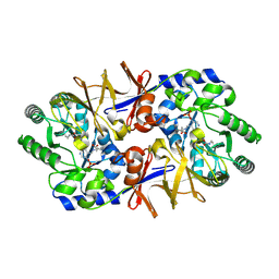
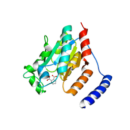
10BT
 
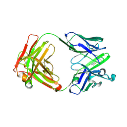 | | X-ray Crystal Structure of A High-Affinity Monoclonal Antibody Sequesters Xylazine | | Descriptor: | Antibody Heavy Chain, Antibody Light Chain, CHLORIDE ION, ... | | Authors: | Shi, K, Moller, N, Aihara, H. | | Deposit date: | 2026-01-10 | | Release date: | 2026-06-10 | | Method: | X-RAY DIFFRACTION (1.99 Å) | | Cite: | Discovery, Structural Characterization, and Preclinical Evaluation of Monoclonal Antibodies against Xylazine Poisoning
Acs Pharmacol Transl Sci, 2026
|
|
10PI
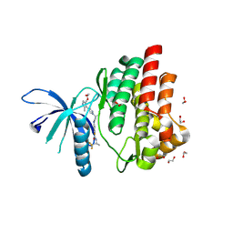 | | JAK1 kinase (JH1 domain) in complex with povorcitinib | | Descriptor: | 1,2-ETHANEDIOL, 4-{3-(cyanomethyl)-3-[(4M)-3',5'-dimethyl-1H,1'H-[4,4'-bipyrazol]-1-yl]azetidin-1-yl}-2,5-difluoro-N-[(2S)-1,1,1-trifluoropropan-2-yl]benzamide, DIMETHYL SULFOXIDE, ... | | Authors: | Epling, L.B, Fenalti, G. | | Deposit date: | 2026-01-30 | | Release date: | 2026-06-10 | | Method: | X-RAY DIFFRACTION (1.54 Å) | | Cite: | Discovery of the Orally Bioavailable Isoform Selective Janus Kinase 1 (JAK1) Compound Povorcitinib (INCB054707) for the Treatment of Inflammatory and Autoimmune Diseases.
J.Med.Chem., 2026
|
|
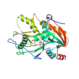
10PJ
 | | JAK2 kinase (JH1 domain) in complex with povorcitinib | | Descriptor: | 4-{3-(cyanomethyl)-3-[(4M)-3',5'-dimethyl-1H,1'H-[4,4'-bipyrazol]-1-yl]azetidin-1-yl}-2,5-difluoro-N-[(2S)-1,1,1-trifluoropropan-2-yl]benzamide, MALONIC ACID, Tyrosine-protein kinase JAK2 | | Authors: | Epling, L.B, Fenalti, G. | | Deposit date: | 2026-01-30 | | Release date: | 2026-06-10 | | Method: | X-RAY DIFFRACTION (1.59 Å) | | Cite: | Discovery of the Orally Bioavailable Isoform Selective Janus Kinase 1 (JAK1) Compound Povorcitinib (INCB054707) for the Treatment of Inflammatory and Autoimmune Diseases.
J.Med.Chem., 2026
|
|
10YZ
 | |
11AO
 | |
11AP
 | |
11UC
 | |
12ZJ
 | |
13LD
 | |
13LV
 | | Structure of PKMYT1 bound to allosteric inhibitor P29-(S) | | Descriptor: | (4S)-1-cyclopentyl-4-(4-hydroxyphenyl)-1,4,5,7-tetrahydro-6H-pyrazolo[3,4-b]pyridin-6-one, Membrane-associated tyrosine- and threonine-specific cdc2-inhibitory kinase | | Authors: | Khamrui, S, Lazarus, M.B. | | Deposit date: | 2026-05-13 | | Release date: | 2026-06-10 | | Method: | X-RAY DIFFRACTION (2.22 Å) | | Cite: | Allosteric Inhibition of PKMYT1 Induces a Unique, Inactive ATP Binding Site Conformation.
J.Am.Chem.Soc., 2026
|
|
13LW
 | | Structure of PKMYT1 bound to inhibitor P32-(S) | | Descriptor: | (4S)-1-cyclopentyl-4-(3-hydroxyphenyl)-1,4,5,7-tetrahydro-6H-pyrazolo[3,4-b]pyridin-6-one, Membrane-associated tyrosine- and threonine-specific cdc2-inhibitory kinase | | Authors: | Khamrui, S, Lazarus, M.B. | | Deposit date: | 2026-05-13 | | Release date: | 2026-06-10 | | Method: | X-RAY DIFFRACTION (1.95 Å) | | Cite: | Allosteric Inhibition of PKMYT1 Induces a Unique, Inactive ATP Binding Site Conformation.
J.Am.Chem.Soc., 2026
|
|
13LX
 | | Structure of PKMYT1 bound to inhibitor P32-(R) | | Descriptor: | (4R)-1-cyclopentyl-4-(3-hydroxyphenyl)-1,4,5,7-tetrahydro-6H-pyrazolo[3,4-b]pyridin-6-one, Membrane-associated tyrosine- and threonine-specific cdc2-inhibitory kinase | | Authors: | Khamrui, S, Lazarus, M.B. | | Deposit date: | 2026-05-13 | | Release date: | 2026-06-10 | | Method: | X-RAY DIFFRACTION (2.2 Å) | | Cite: | Allosteric Inhibition of PKMYT1 Induces a Unique, Inactive ATP Binding Site Conformation.
J.Am.Chem.Soc., 2026
|
|
20ZQ
 | | Crystal structure of rice HPPD | | Descriptor: | 4-hydroxyphenylpyruvate dioxygenase, DI(HYDROXYETHYL)ETHER, GLYCEROL, ... | | Authors: | Chen, L, Ran, T, Wang, W.W, Zhang, B.L. | | Deposit date: | 2025-12-03 | | Release date: | 2026-06-10 | | Method: | X-RAY DIFFRACTION (1.9 Å) | | Cite: | structure of rice hydroxyphenylpyruvate dioxygenase
To Be Published
|
|
21HO
 | | Crystal strucrue of HuHF-C2-MEO complex | | Descriptor: | CHLORIDE ION, Ferritin heavy chain, N-terminally processed, ... | | Authors: | Gong, W.J, Wang, W.M, Wang, H.F. | | Deposit date: | 2025-12-12 | | Release date: | 2026-06-10 | | Method: | X-RAY DIFFRACTION (2.4 Å) | | Cite: | Redesign of the Ferritin Ferroxidase Center for Universal Molecular Binding or Specific Recognition.
Small, 2026
|
|
21KV
 | | Crystal strucrue of HuHF-C2-DAC complex | | Descriptor: | 4-(dimethylaminodiazenyl)-1H-imidazole-5-carboxamide, CALCIUM ION, Ferritin heavy chain, ... | | Authors: | Wang, W.M, Yao, H, Gong, W.J, Wang, H.F. | | Deposit date: | 2025-12-17 | | Release date: | 2026-06-10 | | Method: | X-RAY DIFFRACTION (2 Å) | | Cite: | Redesign of the Ferritin Ferroxidase Center for Universal Molecular Binding or Specific Recognition.
Small, 2026
|
|
21KW
 | | Crystal strucrue of HuHF-C2-SEM complex | | Descriptor: | 1-(2-chloroethyl)-3-(4-methylcyclohexyl)-1-nitroso-urea, CALCIUM ION, Ferritin heavy chain, ... | | Authors: | Wang, W.M, Yao, H, Gong, W.J, Wang, H.F. | | Deposit date: | 2025-12-17 | | Release date: | 2026-06-10 | | Method: | X-RAY DIFFRACTION (2 Å) | | Cite: | Redesign of the Ferritin Ferroxidase Center for Universal Molecular Binding or Specific Recognition.
Small, 2026
|
|
21LE
 | | Crystal strucrue of HuHF-C2-CAR complex | | Descriptor: | CALCIUM ION, CARMUSTINE, Ferritin heavy chain, ... | | Authors: | Wang, W.M, Yao, H, Gong, W.J, Wang, H.F. | | Deposit date: | 2025-12-17 | | Release date: | 2026-06-10 | | Method: | X-RAY DIFFRACTION (2 Å) | | Cite: | Redesign of the Ferritin Ferroxidase Center for Universal Molecular Binding or Specific Recognition.
Small, 2026
|
|
24JD
 | |
24KU
 | |
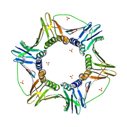
25YR
 | | High-resolution crystal structure of Arp2/3 complex inhibitor Arpin | | Descriptor: | Arpin | | Authors: | Liu, L, Zhang, X, Zhu, Y, Zhang, R, Wang, M, Ye, S. | | Deposit date: | 2026-04-23 | | Release date: | 2026-06-10 | | Method: | X-RAY DIFFRACTION (1.65 Å) | | Cite: | Structural basis of arpin homodimerization reveals cooperative inhibition of the Arp2/3 complex through dual-site engagement.
Protein Cell, 2026
|
|
26QV
 | |
26ST
 | | Crystal structure of Cysteine-dependent hydrolase (CsdH) from Rhodococcus opacus in complex with Monobutylphthalate (MBP) | | Descriptor: | N-carbamoylsarcosine amidohydrolase, PHTHALIC ACID | | Authors: | Aggarwal, S, Singh, S, Jangid, K, Aggarwal, D, Sharma, A.K, Kumar, P. | | Deposit date: | 2026-05-13 | | Release date: | 2026-06-10 | | Method: | X-RAY DIFFRACTION (2.9 Å) | | Cite: | Crystal structure of Cysteine-dependent hydrolases (CsdH) from Rhodococcus opacus
To Be Published
|
|
26ZA
 | | Crystal structure of Cysteine-dependent hydrolase (CsdH) from Rhodococcus opacus in complex with propylene glycol | | Descriptor: | N-carbamoylsarcosine amidohydrolase, S-1,2-PROPANEDIOL | | Authors: | Aggarwal, S, Singh, S, Aggarwal, D, Sharma, A.K, Kumar, P. | | Deposit date: | 2026-05-21 | | Release date: | 2026-06-10 | | Method: | X-RAY DIFFRACTION (2.8 Å) | | Cite: | Crystal structure of Cysteine-dependent hydrolases (CsdH) from Rhodococcus opacus
To Be Published
|
|
27CJ
 | | Crystal structure of Cysteine-dependent hydrolase (CsdH) from Rhodococcus opacus in complex with dibutylphthalate | | Descriptor: | ETHANOL, N-carbamoylsarcosine amidohydrolase, PHTHALIC ACID | | Authors: | Aggarwal, S, Singh, S, Aggarwal, D, Sharma, A.K, Kumar, P. | | Deposit date: | 2026-05-26 | | Release date: | 2026-06-10 | | Method: | X-RAY DIFFRACTION (2.9 Å) | | Cite: | Crystal structure of Cysteine-dependent hydrolase (CsdH) from Rhodococcus opacus in complex with dibutylphthalate
To Be Published
|
|
28JY
 | | Iron loaded E61A human H-chain ferritin, anaerobic | | Descriptor: | CHLORIDE ION, FE (III) ION, Ferritin heavy chain, ... | | Authors: | Bugg, Z, Bradley, J.M, Le Brun, N.E, Hemmings, A.M. | | Deposit date: | 2026-02-04 | | Release date: | 2026-06-10 | | Method: | X-RAY DIFFRACTION (1.6 Å) | | Cite: | Ferritin Iron Mineralisation: Route of Fe 3+ Transfer From the Ferroxidase Centre to the Inner Cavity of Human H-Chain Ferritin.
Angew.Chem.Int.Ed.Engl., 2026
|
|


