 Movie
Movie Controller

[English] 日本語
 Yorodumi
- PDB-5nbz: Wzz dodecamer fitted by MDFF to the Wzz experimental map from cryo-EM -
+ Open data

- Basic information
| Entry | Database: PDB / ID: 5nbz | ||||||
|---|---|---|---|---|---|---|---|
| Title | Wzz dodecamer fitted by MDFF to the Wzz experimental map from cryo-EM | ||||||
 Components Components | WzzB | ||||||
 Keywords Keywords |  MEMBRANE PROTEIN / Wzz Polysaccharide Chain Length membrane protein Cryo-electron microscopy Single particle analysis MEMBRANE PROTEIN / Wzz Polysaccharide Chain Length membrane protein Cryo-electron microscopy Single particle analysis | ||||||
| Function / homology | Polysaccharide chain length determinant N-terminal domain / Chain length determinant protein / lipopolysaccharide biosynthetic process / membrane => GO:0016020 / plasma membrane / Chain length determinant protein Function and homology information Function and homology information | ||||||
| Biological species |   Staphylococcus aureus (bacteria) Staphylococcus aureus (bacteria) | ||||||
| Method | ELECTRON MICROSCOPY / single particle reconstruction / cryo EM / Resolution: 9 Å | ||||||
 Authors Authors | Ford, R.C. / Kargas, V. / Collins, R.F. / Whitfield, C. / Clarke, B.R. / Siebert, A. / Bond, P.J. / Clare, D.K. | ||||||
 Citation Citation | Journal: Structure / Year: 2017 Title: Full-length, Oligomeric Structure of Wzz Determined by Cryoelectron Microscopy Reveals Insights into Membrane-Bound States. Authors: Richard F Collins / Vasileios Kargas / Brad R Clarke / C Alistair Siebert / Daniel K Clare / Peter J Bond / Chris Whitfield / Robert C Ford /    Abstract: Wzz is an integral inner membrane protein involved in regulating the length of lipopolysaccharide O-antigen glycans and essential for the virulence of many Gram-negative pathogens. In all Wzz ...Wzz is an integral inner membrane protein involved in regulating the length of lipopolysaccharide O-antigen glycans and essential for the virulence of many Gram-negative pathogens. In all Wzz homologs, the large periplasmic domain is proposed to be anchored by two transmembrane helices, but no information is available for the transmembrane and cytosolic domains. Here we have studied purified oligomeric Wzz complexes using cryoelectron microscopy and resolved the transmembrane regions within a semi-continuous detergent micelle. The transmembrane helices of each monomer display a right-handed super-helical twist, and do not interact with the neighboring transmembrane domains. Modeling, flexible fitting and multiscale simulation approaches were used to study the full-length complex and to provide explanations for the influence of the lipid bilayer on its oligomeric status. Based on structural and in silico observations, we propose a new mechanism for O-antigen chain-length regulation that invokes synergy of Wzz and its polymerase partner, Wzy. | ||||||
| History |
|
- Structure visualization
| Movie |
Movie viewer |
|---|---|
| Structure viewer | Molecule: MolmilJmol/JSmol |
- Downloads & links
-Download
| PDBx/mmCIF format | 5nbz.cif.gz | 908.3 KB | Display | PDBx/mmCIF format |
|---|---|---|---|---|
| PDB format | pdb5nbz.ent.gz | 770 KB | Display | PDB format |
| PDBx/mmJSON format | 5nbz.json.gz | Tree view | PDBx/mmJSON format | |
| Others |  Other downloads Other downloads |
-Validation report
| Arichive directory | https://data.pdbj.org/pub/pdb/validation_reports/nb/5nbzftp://data.pdbj.org/pub/pdb/validation_reports/nb/5nbz | HTTPS FTP |
|---|
-Related structure data
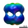
| Related structure data | 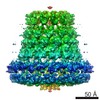 3611MC M: map data used to model this data C: citing same article ( |
|---|---|
| Similar structure data |


-Links
 PDBj
PDBj- Assembly
| Deposited unit | 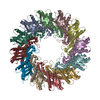
|
|---|---|
| 1 |
|
-Components
| #1: Protein | Mass: 26760.129 Da / Num. of mol.: 12 Source method: isolated from a genetically manipulated source Source: (gene. exp.) Staphylococcus aureus (bacteria) / Production host: Escherichia coli (E. coli) / References: UniProt: P35272*PLUS |
|---|
-Experimental details
-Experiment
| Experiment | Method: ELECTRON MICROSCOPY |
|---|---|
| EM experiment | Aggregation state: PARTICLE / 3D reconstruction method: single particle reconstruction |
- Sample preparation
| Component | Name: Dodecameric complex / Type: COMPLEX / Entity ID: all / Source: RECOMBINANT |
|---|---|
| Molecular weight | Value: 0.36 MDa / Experimental value: NO |
| Source (natural) | Organism: Staphylococcus aureus (bacteria) |
| Source (recombinant) | Organism: Escherichia coli (E. coli) |
| Buffer solution | pH: 7.5 Details: 20mM Tris pH 7.5, 150mM NaCl, 0.025% dodecyl maltoside. |
| Specimen | Conc.: 0.3 mg/ml / Embedding applied: NO / Shadowing applied: NO / Staining applied: NO / Vitrification applied: YES Details: Dodecamers with C12 symmetry with a small fraction of head-to-head D12 complexes. |
| Specimen support | Grid material: GOLD / Grid mesh size: 400 divisions/in. / Grid type: Quantifoil R1.2/1.3 |
| Vitrification | Instrument: FEI VITROBOT MARK III / Cryogen name: ETHANE / Humidity: 95 % / Chamber temperature: 293 K / Details: 1 x 4sec blot |
- Electron microscopy imaging
| Experimental equipment |  Model: Titan Krios / Image courtesy: FEI Company |
|---|---|
| Microscopy | Model: FEI TITAN KRIOS |
| Electron gun | Electron source: FIELD EMISSION GUN / Accelerating voltage: 300 kV / Illumination mode: FLOOD BEAM |
| Electron lens | Mode: BRIGHT FIELDBright-field microscopy |
| Specimen holder | Cryogen: NITROGEN / Specimen holder model: FEI TITAN KRIOS AUTOGRID HOLDER |
| Image recording | Electron dose: 2 e/Å2 / Film or detector model: GATAN K2 SUMMIT (4k x 4k) |
- Processing
| CTF correction | Type: PHASE FLIPPING AND AMPLITUDE CORRECTION |
|---|---|
| 3D reconstruction | Resolution: 9 Å / Resolution method: OTHER / Num. of particles: 22000 / Details: ResMap / Symmetry type: POINT |
| Atomic model building | Protocol: FLEXIBLE FIT / Space: REAL |
| Atomic model building | PDB-ID: 4.0E+29 / Pdb chain-ID: A / Pdb chain residue range: 55-291 |
