 Movie
Movie Controller


+ Open data

- Basic information
| Entry | Database: PDB / ID: 3j3r | ||||||
|---|---|---|---|---|---|---|---|
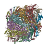
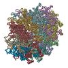
| Title | Structural dynamics of the MecA-ClpC complex revealed by cryo-EM | ||||||
 Components Components |
| ||||||
 Keywords Keywords | CHAPERONE / ClpC / MecA / AAA+ ATPase / PROTEIN unfolding | ||||||
| Function / homology |  Function and homology information Function and homology informationnegative regulation of establishment of competence for transformation / negative regulation of sporulation resulting in formation of a cellular spore / establishment of competence for transformation / sporulation resulting in formation of a cellular spore / protein folding chaperone / protein-macromolecule adaptor activity / ATP hydrolysis activity / ATP binding Similarity search - Function | ||||||
| Biological species |  | ||||||
| Method | ELECTRON MICROSCOPY / single particle reconstruction / cryo EM / Resolution: 9.4 Å | ||||||
 Authors Authors | Liu, J. / Mei, Z. / Li, N. / Qi, Y. / Xu, Y. / Shi, Y. / Wang, F. / Lei, J. / Gao, N. | ||||||
 Citation Citation | Journal: J Biol Chem / Year: 2013 Title: Structural dynamics of the MecA-ClpC complex: a type II AAA+ protein unfolding machine. Authors: Jing Liu / Ziqing Mei / Ningning Li / Yutao Qi / Yanji Xu / Yigong Shi / Feng Wang / Jianlin Lei / Ning Gao /  Abstract: The MecA-ClpC complex is a bacterial type II AAA(+) molecular machine responsible for regulated unfolding of substrates, such as transcription factors ComK and ComS, and targeting them to ClpP for ...The MecA-ClpC complex is a bacterial type II AAA(+) molecular machine responsible for regulated unfolding of substrates, such as transcription factors ComK and ComS, and targeting them to ClpP for degradation. The six subunits of the MecA-ClpC complex form a closed barrel-like structure, featured with three stacked rings and a hollow passage, where substrates are threaded and translocated through successive pores. Although the general concepts of how polypeptides are unfolded and translocated by internal pore loops of AAA(+) proteins have long been conceived, the detailed mechanistic model remains elusive. With cryoelectron microscopy, we captured four different structures of the MecA-ClpC complexes. These complexes differ in the nucleotide binding states of the two AAA(+) rings and therefore might presumably reflect distinctive, representative snapshots from a dynamic unfolding cycle of this hexameric complex. Structural analysis reveals that nucleotide binding and hydrolysis modulate the hexameric complex in a number of ways, including the opening of the N-terminal ring, the axial and radial positions of pore loops, the compactness of the C-terminal ring, as well as the relative rotation between the two nucleotide-binding domain rings. More importantly, our structural and biochemical data indicate there is an active allosteric communication between the two AAA(+) rings and suggest that concerted actions of the two AAA(+) rings are required for the efficiency of the substrate unfolding and translocation. These findings provide important mechanistic insights into the dynamic cycle of the MecA-ClpC unfoldase and especially lay a foundation toward the complete understanding of the structural dynamics of the general type II AAA(+) hexamers. | ||||||
| History |
|
- Structure visualization
| Movie |
Movie viewer |
|---|---|
| Structure viewer | Molecule: MolmilJmol/JSmol |
- Downloads & links
-Download
| PDBx/mmCIF format | 3j3r.cif.gz | 889.3 KB | Display | PDBx/mmCIF format |
|---|---|---|---|---|
| PDB format | pdb3j3r.ent.gz | 710.1 KB | Display | PDB format |
| PDBx/mmJSON format | 3j3r.json.gz | Tree view | PDBx/mmJSON format | |
| Others |  Other downloads Other downloads |
-Validation report
| Arichive directory | https://data.pdbj.org/pub/pdb/validation_reports/j3/3j3rftp://data.pdbj.org/pub/pdb/validation_reports/j3/3j3r | HTTPS FTP |
|---|
-Related structure data
| Related structure data |  5610MC  5607C 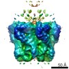 5608C 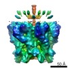 5609C  3j3sC  3j3tC  3j3uC M: map data used to model this data C: citing same article ( |
|---|---|
| Similar structure data |


-Links
 PDBj
PDBj


- Assembly
| Deposited unit | 
|
|---|---|
| 1 |
|
-Components
| #1: Protein | Mass: 25791.617 Da / Num. of mol.: 6 Source method: isolated from a genetically manipulated source Source: (gene. exp.) #2: Protein | Mass: 90135.648 Da / Num. of mol.: 6 / Mutation: E280A, E618A Source method: isolated from a genetically manipulated source Source: (gene. exp.) |
|---|
-Experimental details
-Experiment
| Experiment | Method: ELECTRON MICROSCOPY |
|---|---|
| EM experiment | Aggregation state: PARTICLE / 3D reconstruction method: single particle reconstruction |
- Sample preparation
| Component | Name: MecA-ClpC (E280A, E618A) with ADP / Type: COMPLEX / Details: hexamer |
|---|---|
| Molecular weight | Value: 0.6 MDa / Experimental value: NO |
| Buffer solution | Name: 50mM kCl, 10mM Tris-HCL, 2mM MgCl2, 2mM ADP / pH: 7.5 / Details: 50mM kCl, 10mM Tris-HCL, 2mM MgCl2, 2mM ADP |
| Specimen | Conc.: 0.03 mg/ml / Embedding applied: NO / Shadowing applied: NO / Staining applied: NO / Vitrification applied: YES |
| Vitrification | Instrument: FEI VITROBOT MARK IV / Cryogen name: ETHANE / Temp: 90 K / Humidity: 100 % / Method: Blot for 2 seconds before plunging |
- Electron microscopy imaging
| Experimental equipment |  Model: Titan Krios / Image courtesy: FEI Company |
|---|---|
| Microscopy | Model: FEI TITAN KRIOS / Date: Sep 9, 2010 |
| Electron gun | Electron source:  FIELD EMISSION GUN / Accelerating voltage: 300 kV / Illumination mode: FLOOD BEAM FIELD EMISSION GUN / Accelerating voltage: 300 kV / Illumination mode: FLOOD BEAM |
| Electron lens | Mode: BRIGHT FIELD / Nominal magnification: 59000 X / Nominal defocus max: 4000 nm / Nominal defocus min: 1500 nm / Camera length: 0 mm |
| Specimen holder | Specimen holder model: FEI TITAN KRIOS AUTOGRID HOLDER / Tilt angle max: 0 ° / Tilt angle min: 0 ° |
| Image recording | Electron dose: 20 e/Å2 / Film or detector model: FEI EAGLE (4k x 4k) |
| Radiation wavelength | Relative weight: 1 |
- Processing
| EM software |
| ||||||||||||
|---|---|---|---|---|---|---|---|---|---|---|---|---|---|
| CTF correction | Details: each defocus group on 3D level | ||||||||||||
| Symmetry | Point symmetry: C1 (asymmetric) | ||||||||||||
| 3D reconstruction | Method: Reference Projections / Resolution: 9.4 Å / Resolution method: FSC 0.5 CUT-OFF / Num. of particles: 26037 / Details: Single particle--Applied symmetry: C6 / Symmetry type: POINT | ||||||||||||
| Atomic model building | Protocol: FLEXIBLE FIT / Space: REAL / Target criteria: Cross-correlation Details: REFINEMENT PROTOCOL--flexible DETAILS--Protocol- Initial local fitting was done using Chimera and then MDFF was used for flexible fitting. ref- Trabuco, L.G., Villa, E., Mitra, K., Frank, J. ...Details: REFINEMENT PROTOCOL--flexible DETAILS--Protocol- Initial local fitting was done using Chimera and then MDFF was used for flexible fitting. ref- Trabuco, L.G., Villa, E., Mitra, K., Frank, J. and Schulten, K. (2008) Flexible fitting of atomic structures into electron microscopy maps using molecular dynamics. | ||||||||||||
| Atomic model building | PDB-ID: 3PXI Accession code: 3PXI / Source name: PDB / Type: experimental model | ||||||||||||
| Refinement step | Cycle: LAST
|

