 Movie
Movie Controller


+ Open data

- Basic information
| Entry | Database: PDB / ID: 6z4u | ||||||
|---|---|---|---|---|---|---|---|
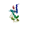
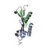
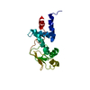
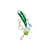
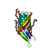
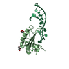
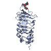
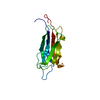
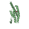
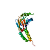
| Title | X-ray Crystallographic Structure of Orf9b from SARS-CoV-2 | ||||||
 Components Components | Protein 9b | ||||||
 Keywords Keywords | VIRAL PROTEIN / orf9b | ||||||
| Function / homology |  Function and homology information Function and homology informationTranslation of Accessory Proteins / outer mitochondrial membrane protein complex / negative regulation of defense response to virus / positive regulation of autophagosome assembly / negative regulation of mitochondrial fission / host cell mitochondrion / protein sequestering activity / symbiont-mediated suppression of host cytoplasmic pattern recognition receptor signaling pathway via inhibition of MAVS activity / DDX58/IFIH1-mediated induction of interferon-alpha/beta / mitochondrial membrane ...Translation of Accessory Proteins / outer mitochondrial membrane protein complex / negative regulation of defense response to virus / positive regulation of autophagosome assembly / negative regulation of mitochondrial fission / host cell mitochondrion / protein sequestering activity / symbiont-mediated suppression of host cytoplasmic pattern recognition receptor signaling pathway via inhibition of MAVS activity / DDX58/IFIH1-mediated induction of interferon-alpha/beta / mitochondrial membrane / mitochondrial outer membrane / symbiont-mediated suppression of host type I interferon-mediated signaling pathway / virus-mediated perturbation of host defense response / SARS-CoV-2 activates/modulates innate and adaptive immune responses / identical protein binding Similarity search - Function | ||||||
| Biological species |   Severe acute respiratory syndrome coronavirus 2 Severe acute respiratory syndrome coronavirus 2 | ||||||
| Method |  X-RAY DIFFRACTION / SYNCHROTRON / MOLECULAR REPLACEMENT / Resolution: 1.95 Å X-RAY DIFFRACTION / SYNCHROTRON / MOLECULAR REPLACEMENT / Resolution: 1.95 Å | ||||||
 Authors Authors | Weeks, S.D. / De Graef, S. / Munawar, A. | ||||||
 Citation Citation | Journal: To Be Published Title: X-ray Crystallographic Structure of Orf9b from SARS-CoV-2 Authors: Weeks, S.D. / De Graef, S. / Munawar, A. | ||||||
| History |
|
- Structure visualization
| Structure viewer | Molecule: MolmilJmol/JSmol |
|---|
- Downloads & links
-Download
| PDBx/mmCIF format | 6z4u.cif.gz | 81 KB | Display | PDBx/mmCIF format |
|---|---|---|---|---|
| PDB format | pdb6z4u.ent.gz | 57.6 KB | Display | PDB format |
| PDBx/mmJSON format | 6z4u.json.gz | Tree view | PDBx/mmJSON format | |
| Others |  Other downloads Other downloads |
-Validation report
| Summary document | 6z4u_validation.pdf.gz | 593 KB | Display | wwPDB validaton report |
|---|---|---|---|---|
| Full document | 6z4u_full_validation.pdf.gz | 593.2 KB | Display | |
| Data in XML | 6z4u_validation.xml.gz | 8.8 KB | Display | |
| Data in CIF | 6z4u_validation.cif.gz | 11.3 KB | Display | |
| Arichive directory | https://data.pdbj.org/pub/pdb/validation_reports/z4/6z4uftp://data.pdbj.org/pub/pdb/validation_reports/z4/6z4u | HTTPS FTP |
-Related structure data
| Related structure data | 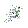 2cmeS S: Starting model for refinement |
|---|---|
| Similar structure data |
-Links
 PDBj
PDBj- Assembly
| Deposited unit | 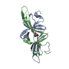
| ||||||||
|---|---|---|---|---|---|---|---|---|---|
| 1 |
| ||||||||
| Unit cell |
|
-Components
| #1: Protein | Mass: 10808.636 Da / Num. of mol.: 2 Source method: isolated from a genetically manipulated source Source: (gene. exp.) Severe acute respiratory syndrome coronavirus 2Production host:  #2: Chemical | ChemComp-15P / |   Mass: 1529.829 Da / Num. of mol.: 1 / Source method: obtained synthetically / Formula: C69H140O35 / Comment: precipitant*YM Mass: 1529.829 Da / Num. of mol.: 1 / Source method: obtained synthetically / Formula: C69H140O35 / Comment: precipitant*YM#3: Water | ChemComp-HOH / |  Mass: 18.015 Da / Num. of mol.: 49 / Source method: isolated from a natural source / Formula: H2O Mass: 18.015 Da / Num. of mol.: 49 / Source method: isolated from a natural source / Formula: H2OHas ligand of interest | N | |
|---|
-Experimental details
-Experiment
| Experiment | Method: X-RAY DIFFRACTION / Number of used crystals: 1 |
|---|
- Sample preparation
| Crystal | Density Matthews: 1.98 Å3/Da / Density % sol: 37.83 % |
|---|---|
| Crystal grow | Temperature: 293.15 K / Method: vapor diffusion, sitting drop / pH: 6.5 Details: Protein at 8.3 mg/ml in 20 mM Tris pH 7.0 150 mM NaCl, 1mM DTT was mixed in a 3:2 ratio with the MORPHEUS crystallization screen condition E4 (Tube 2-4; 0.12 M Ethylene glycols 0.1 M Buffer ...Details: Protein at 8.3 mg/ml in 20 mM Tris pH 7.0 150 mM NaCl, 1mM DTT was mixed in a 3:2 ratio with the MORPHEUS crystallization screen condition E4 (Tube 2-4; 0.12 M Ethylene glycols 0.1 M Buffer System 1 pH 6.5 37.5 % v/v Precipitant Mix 4) |
-Data collection
| Diffraction | Mean temperature: 100 K / Serial crystal experiment: N | ||||||||||||||||||||||||
|---|---|---|---|---|---|---|---|---|---|---|---|---|---|---|---|---|---|---|---|---|---|---|---|---|---|
| Diffraction source | Source: SYNCHROTRON / Site: NSLS-II  / Beamline: 17-ID-2 / Wavelength: 0.979329 Å / Beamline: 17-ID-2 / Wavelength: 0.979329 Å | ||||||||||||||||||||||||
| Detector | Type: DECTRIS EIGER X 16M / Detector: PIXEL / Date: May 15, 2020 | ||||||||||||||||||||||||
| Radiation | Monochromator: Si 111 double crystal / Protocol: SINGLE WAVELENGTH / Monochromatic (M) / Laue (L): M / Scattering type: x-ray | ||||||||||||||||||||||||
| Radiation wavelength | Wavelength: 0.979329 Å / Relative weight: 1 | ||||||||||||||||||||||||
| Reflection | Resolution: 1.95→36.77 Å / Num. obs: 13064 / % possible obs: 99.3 % / Redundancy: 7.4 % / CC1/2: 0.934 / Rmerge(I) obs: 0.123 / Rpim(I) all: 0.05 / Rrim(I) all: 0.133 / Net I/σ(I): 8.5 | ||||||||||||||||||||||||
| Reflection shell | Diffraction-ID: 1
|
- Processing
| Software |
| |||||||||||||||||||||||||||||||||||||||||||||||||||||||||||||||||||||||||||
|---|---|---|---|---|---|---|---|---|---|---|---|---|---|---|---|---|---|---|---|---|---|---|---|---|---|---|---|---|---|---|---|---|---|---|---|---|---|---|---|---|---|---|---|---|---|---|---|---|---|---|---|---|---|---|---|---|---|---|---|---|---|---|---|---|---|---|---|---|---|---|---|---|---|---|---|---|
| Refinement | Method to determine structure: MOLECULAR REPLACEMENT Starting model: 2CME Resolution: 1.95→36.77 Å / Cor.coef. Fo:Fc: 0.945 / Cor.coef. Fo:Fc free: 0.922 / SU B: 9.017 / SU ML: 0.13 / Cross valid method: THROUGHOUT / σ(F): 0 / ESU R: 0.186 / ESU R Free: 0.166 / Stereochemistry target values: MAXIMUM LIKELIHOOD / Details: U VALUES : WITH TLS ADDED
| |||||||||||||||||||||||||||||||||||||||||||||||||||||||||||||||||||||||||||
| Solvent computation | Ion probe radii: 0.8 Å / Shrinkage radii: 0.8 Å / VDW probe radii: 1.2 Å / Solvent model: MASK | |||||||||||||||||||||||||||||||||||||||||||||||||||||||||||||||||||||||||||
| Displacement parameters | Biso max: 82.54 Å2 / Biso mean: 33.585 Å2 / Biso min: 16.12 Å2
| |||||||||||||||||||||||||||||||||||||||||||||||||||||||||||||||||||||||||||
| Refinement step | Cycle: final / Resolution: 1.95→36.77 Å
| |||||||||||||||||||||||||||||||||||||||||||||||||||||||||||||||||||||||||||
| Refine LS restraints |
| |||||||||||||||||||||||||||||||||||||||||||||||||||||||||||||||||||||||||||
| LS refinement shell | Resolution: 1.95→2.001 Å / Rfactor Rfree error: 0 / Total num. of bins used: 20
| |||||||||||||||||||||||||||||||||||||||||||||||||||||||||||||||||||||||||||
| Refinement TLS params. | Method: refined / Refine-ID: X-RAY DIFFRACTION
| |||||||||||||||||||||||||||||||||||||||||||||||||||||||||||||||||||||||||||
| Refinement TLS group |
|
