 Movie
Movie Controller

[English] 日本語
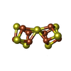
 Yorodumi
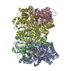
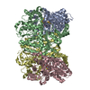
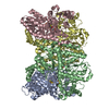
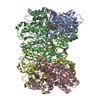
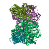
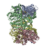
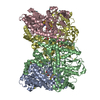
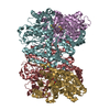
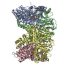
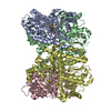
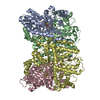
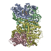
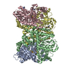
- PDB-6o7r: Nitrogenase MoFeP mutant F99Y, S188A from Azotobacter vinelandii ... -
+ Open data

- Basic information
| Entry | Database: PDB / ID: 6o7r | |||||||||
|---|---|---|---|---|---|---|---|---|---|---|
| Title | Nitrogenase MoFeP mutant F99Y, S188A from Azotobacter vinelandii in the dithionite reduced state | |||||||||
 Components Components | (Nitrogenase molybdenum-iron protein ...) x 2 | |||||||||
 Keywords Keywords | OXIDOREDUCTASE / Nitrogenase / F99Y/S188A / MoFeP | |||||||||
| Function / homology |  Function and homology information Function and homology informationmolybdenum-iron nitrogenase complex / nitrogenase / nitrogenase activity / nitrogen fixation / iron-sulfur cluster binding / ATP binding / metal ion binding Similarity search - Function | |||||||||
| Biological species |  Azotobacter vinelandii (bacteria) Azotobacter vinelandii (bacteria) | |||||||||
| Method |  X-RAY DIFFRACTION / SYNCHROTRON / MOLECULAR REPLACEMENT / Resolution: 2.27 Å X-RAY DIFFRACTION / SYNCHROTRON / MOLECULAR REPLACEMENT / Resolution: 2.27 Å | |||||||||
 Authors Authors | Rutledge, H.L. / Williamson, L.M. / Tezcan, F.A. | |||||||||
| Funding support |  United States, 2items United States, 2items
| |||||||||
 Citation Citation | Journal: J.Am.Chem.Soc. / Year: 2019 Title: Redox-Dependent Metastability of the Nitrogenase P-Cluster. Authors: Rutledge, H.L. / Rittle, J. / Williamson, L.M. / Xu, W.A. / Gagnon, D.M. / Tezcan, F.A. | |||||||||
| History |
|
- Structure visualization
| Structure viewer | Molecule: MolmilJmol/JSmol |
|---|
- Downloads & links
-Download
| PDBx/mmCIF format | 6o7r.cif.gz | 758.3 KB | Display | PDBx/mmCIF format |
|---|---|---|---|---|
| PDB format | pdb6o7r.ent.gz | 625.6 KB | Display | PDB format |
| PDBx/mmJSON format | 6o7r.json.gz | Tree view | PDBx/mmJSON format | |
| Others |  Other downloads Other downloads |
-Validation report
| Arichive directory | https://data.pdbj.org/pub/pdb/validation_reports/o7/6o7rftp://data.pdbj.org/pub/pdb/validation_reports/o7/6o7r | HTTPS FTP |
|---|
-Related structure data
| Related structure data | 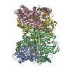 6o7lC  6o7mC  6o7nC  6o7oC 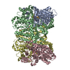 6o7pC  6o7qC 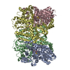 6o7sC 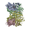 2minS S: Starting model for refinement C: citing same article ( |
|---|---|
| Similar structure data |


-Links
 PDBj
PDBj
- Assembly
| Deposited unit | 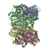
| ||||||||
|---|---|---|---|---|---|---|---|---|---|
| 1 |
| ||||||||
| Unit cell |
|
-Components
-Nitrogenase molybdenum-iron protein ... , 2 types, 4 molecules ACBD
| #1: Protein | Mass: 55363.043 Da / Num. of mol.: 2 / Source method: isolated from a natural source / Details: nifD / Source: (natural) Azotobacter vinelandii (bacteria) / References: UniProt: P07328, nitrogenase#2: Protein | Mass: 59535.879 Da / Num. of mol.: 2 / Mutation: F99Y, S188A / Source method: isolated from a natural source / Details: nifD / Source: (natural) Azotobacter vinelandii (bacteria) / References: UniProt: P07329, nitrogenase |
|---|
-Non-polymers , 5 types, 1335 molecules 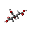
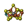


| #3: Chemical |  Mass: 206.150 Da / Num. of mol.: 2 / Source method: obtained synthetically / Formula: C7H10O7 Mass: 206.150 Da / Num. of mol.: 2 / Source method: obtained synthetically / Formula: C7H10O7#4: Chemical |  Mass: 787.451 Da / Num. of mol.: 2 / Source method: obtained synthetically / Formula: CFe7MoS9 Mass: 787.451 Da / Num. of mol.: 2 / Source method: obtained synthetically / Formula: CFe7MoS9#5: Chemical |  Mass: 671.215 Da / Num. of mol.: 2 / Source method: obtained synthetically / Formula: Fe8S7 / Feature type: SUBJECT OF INVESTIGATION Mass: 671.215 Da / Num. of mol.: 2 / Source method: obtained synthetically / Formula: Fe8S7 / Feature type: SUBJECT OF INVESTIGATION#6: Chemical |  Mass: 55.845 Da / Num. of mol.: 2 / Source method: obtained synthetically / Formula: Fe Mass: 55.845 Da / Num. of mol.: 2 / Source method: obtained synthetically / Formula: Fe#7: Water | ChemComp-HOH / | Mass: 18.015 Da / Num. of mol.: 1327 / Source method: isolated from a natural source / Formula: H2O |
|---|
-Experimental details
-Experiment
| Experiment | Method: X-RAY DIFFRACTION / Number of used crystals: 1 |
|---|
- Sample preparation
| Crystal | Density Matthews: 2.18 Å3/Da / Density % sol: 43.69 % |
|---|---|
| Crystal grow | Temperature: 293 K / Method: vapor diffusion, sitting drop / pH: 8 Details: 20% PEG 8000, 100 mM Tris pH 8.0, 500 mM NaCl, 10 mM dithionite |
-Data collection
| Diffraction | Mean temperature: 100 K / Serial crystal experiment: N |
|---|---|
| Diffraction source | Source: SYNCHROTRON / Site: SSRL / Beamline: BL9-2 / Wavelength: 1.7388 Å |
| Detector | Type: DECTRIS PILATUS 6M / Detector: PIXEL / Date: Jun 29, 2018 / Details: Rh coated flat bent M0 |
| Radiation | Monochromator: Si(111) and Si(220) double crystal / Protocol: SINGLE WAVELENGTH / Monochromatic (M) / Laue (L): M / Scattering type: x-ray |
| Radiation wavelength | Wavelength: 1.7388 Å / Relative weight: 1 |
| Reflection | Resolution: 2.27→79.84 Å / Num. obs: 82483 / % possible obs: 90.7 % / Redundancy: 5.8 % / CC1/2: 0.984 / Rmerge(I) obs: 0.114 / Rpim(I) all: 0.079 / Rrim(I) all: 0.14 / Χ2: 0.99 / Net I/σ(I): 9.7 |
| Reflection shell | Resolution: 2.27→2.31 Å / Redundancy: 5.6 % / Rmerge(I) obs: 0.383 / Mean I/σ(I) obs: 3.8 / Num. unique obs: 4590 / CC1/2: 0.901 / Rpim(I) all: 0.269 / Rrim(I) all: 0.471 / Χ2: 0.96 / % possible all: 95.3 |
- Processing
| Software |
| |||||||||||||||||||||||||||||||||||||||||||||||||||||||||||||||||||||||||||||||||||||||||||||||||||||||||||||||||||||||||||||||||||||||||||||||||||||||||||||||||||||||||||||||||||||||||||||||||||||||||||||||||||||||||
|---|---|---|---|---|---|---|---|---|---|---|---|---|---|---|---|---|---|---|---|---|---|---|---|---|---|---|---|---|---|---|---|---|---|---|---|---|---|---|---|---|---|---|---|---|---|---|---|---|---|---|---|---|---|---|---|---|---|---|---|---|---|---|---|---|---|---|---|---|---|---|---|---|---|---|---|---|---|---|---|---|---|---|---|---|---|---|---|---|---|---|---|---|---|---|---|---|---|---|---|---|---|---|---|---|---|---|---|---|---|---|---|---|---|---|---|---|---|---|---|---|---|---|---|---|---|---|---|---|---|---|---|---|---|---|---|---|---|---|---|---|---|---|---|---|---|---|---|---|---|---|---|---|---|---|---|---|---|---|---|---|---|---|---|---|---|---|---|---|---|---|---|---|---|---|---|---|---|---|---|---|---|---|---|---|---|---|---|---|---|---|---|---|---|---|---|---|---|---|---|---|---|---|---|---|---|---|---|---|---|---|---|---|---|---|---|---|---|---|
| Refinement | Method to determine structure: MOLECULAR REPLACEMENT Starting model: 2MIN Resolution: 2.27→47.311 Å / SU ML: 0.22 / Cross valid method: FREE R-VALUE / σ(F): 1.34 / Phase error: 20.51
| |||||||||||||||||||||||||||||||||||||||||||||||||||||||||||||||||||||||||||||||||||||||||||||||||||||||||||||||||||||||||||||||||||||||||||||||||||||||||||||||||||||||||||||||||||||||||||||||||||||||||||||||||||||||||
| Solvent computation | Shrinkage radii: 0.9 Å / VDW probe radii: 1.11 Å | |||||||||||||||||||||||||||||||||||||||||||||||||||||||||||||||||||||||||||||||||||||||||||||||||||||||||||||||||||||||||||||||||||||||||||||||||||||||||||||||||||||||||||||||||||||||||||||||||||||||||||||||||||||||||
| Refinement step | Cycle: LAST / Resolution: 2.27→47.311 Å
| |||||||||||||||||||||||||||||||||||||||||||||||||||||||||||||||||||||||||||||||||||||||||||||||||||||||||||||||||||||||||||||||||||||||||||||||||||||||||||||||||||||||||||||||||||||||||||||||||||||||||||||||||||||||||
| Refine LS restraints |
| |||||||||||||||||||||||||||||||||||||||||||||||||||||||||||||||||||||||||||||||||||||||||||||||||||||||||||||||||||||||||||||||||||||||||||||||||||||||||||||||||||||||||||||||||||||||||||||||||||||||||||||||||||||||||
| LS refinement shell |
|
