 Movie
Movie Controller

[English] 日本語
 Yorodumi
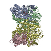
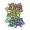
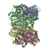
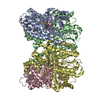
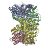
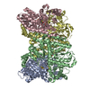
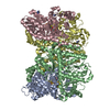
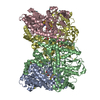
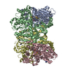
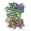
- PDB-2min: NITROGENASE MOFE PROTEIN FROM AZOTOBACTER VINELANDII, OXIDIZED STATE -
+ Open data

- Basic information
| Entry | Database: PDB / ID: 2min | |||||||||
|---|---|---|---|---|---|---|---|---|---|---|
| Title | NITROGENASE MOFE PROTEIN FROM AZOTOBACTER VINELANDII, OXIDIZED STATE | |||||||||
 Components Components | (NITROGENASE MOLYBDENUM IRON ...) x 2 | |||||||||
 Keywords Keywords | NITROGEN FIXATION / NITROGEN METABOLISM / OXIDOREDUCTASE / MOLYBDOENZYMES / BIOLOGICAL NITROGEN FIXATION | |||||||||
| Function / homology |  Function and homology information Function and homology informationnitrogen fixation / molybdenum-iron nitrogenase complex / nitrogenase / nitrogenase activity / iron-sulfur cluster binding / ATP binding / metal ion binding Similarity search - Function | |||||||||
| Biological species |  Azotobacter vinelandii (bacteria) Azotobacter vinelandii (bacteria) | |||||||||
| Method |  X-RAY DIFFRACTION / SYNCHROTRON / Resolution: 2.03 Å X-RAY DIFFRACTION / SYNCHROTRON / Resolution: 2.03 Å | |||||||||
 Authors Authors | Peters, J.W. / Stowell, M.H.B. / Soltis, S.M. / Day, M.W. / Kim, J. / Rees, D.C. | |||||||||
 Citation Citation | Journal: Biochemistry / Year: 1997 Title: Redox-dependent structural changes in the nitrogenase P-cluster. Authors: Peters, J.W. / Stowell, M.H. / Soltis, S.M. / Finnegan, M.G. / Johnson, M.K. / Rees, D.C. #1: Journal: Nature / Year: 1992Title: Crystallographic Structure and Functional Implications of the Nitrogenase Molybdenum-Iron Protein from Azotobacter Vinelandii Authors: Kim, J. / Rees, D.C. #2: Journal: Science / Year: 1992Title: Structural Models for the Metal Centers in the Nitrogenase Molybdenum-Iron Protein Authors: Kim, J. / Rees, D.C. | |||||||||
| History |
|
- Structure visualization
| Structure viewer | Molecule: MolmilJmol/JSmol |
|---|
- Downloads & links
-Download
| PDBx/mmCIF format | 2min.cif.gz | 415.5 KB | Display | PDBx/mmCIF format |
|---|---|---|---|---|
| PDB format | pdb2min.ent.gz | 331 KB | Display | PDB format |
| PDBx/mmJSON format | 2min.json.gz | Tree view | PDBx/mmJSON format | |
| Others |  Other downloads Other downloads |
-Validation report
| Arichive directory | https://data.pdbj.org/pub/pdb/validation_reports/mi/2minftp://data.pdbj.org/pub/pdb/validation_reports/mi/2min | HTTPS FTP |
|---|
-Related structure data


-Links
 PDBj
PDBj
- Assembly
| Deposited unit | 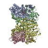
| ||||||||||||
|---|---|---|---|---|---|---|---|---|---|---|---|---|---|
| 1 |
| ||||||||||||
| Unit cell |
| ||||||||||||
| Noncrystallographic symmetry (NCS) | NCS oper:
|
-Components
-NITROGENASE MOLYBDENUM IRON ... , 2 types, 4 molecules ACBD
| #1: Protein | Mass: 55231.848 Da / Num. of mol.: 2 / Source method: isolated from a natural source / Source: (natural) Azotobacter vinelandii (bacteria) / Strain: WILD-TYPE / References: UniProt: P07328, nitrogenase#2: Protein | Mass: 59404.684 Da / Num. of mol.: 2 / Source method: isolated from a natural source / Source: (natural) Azotobacter vinelandii (bacteria) / Strain: WILD-TYPE / References: UniProt: P07329, nitrogenase |
|---|
-Non-polymers , 5 types, 635 molecules 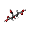
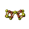
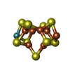


| #3: Chemical |  Mass: 206.150 Da / Num. of mol.: 2 / Source method: obtained synthetically / Formula: C7H10O7 Mass: 206.150 Da / Num. of mol.: 2 / Source method: obtained synthetically / Formula: C7H10O7#4: Chemical |  Mass: 671.215 Da / Num. of mol.: 2 / Source method: obtained synthetically / Formula: Fe8S7 Mass: 671.215 Da / Num. of mol.: 2 / Source method: obtained synthetically / Formula: Fe8S7#5: Chemical |  Mass: 775.440 Da / Num. of mol.: 2 / Source method: obtained synthetically / Formula: Fe7MoS9 Mass: 775.440 Da / Num. of mol.: 2 / Source method: obtained synthetically / Formula: Fe7MoS9#6: Chemical |  Mass: 40.078 Da / Num. of mol.: 2 / Source method: obtained synthetically / Formula: Ca Mass: 40.078 Da / Num. of mol.: 2 / Source method: obtained synthetically / Formula: Ca#7: Water | ChemComp-HOH / | Mass: 18.015 Da / Num. of mol.: 627 / Source method: isolated from a natural source / Formula: H2O |
|---|
-Experimental details
-Experiment
| Experiment | Method: X-RAY DIFFRACTION / Number of used crystals: 1 |
|---|
- Sample preparation
| Crystal | Density Matthews: 2.4 Å3/Da / Density % sol: 49 % | ||||||||||||||||||||||||
|---|---|---|---|---|---|---|---|---|---|---|---|---|---|---|---|---|---|---|---|---|---|---|---|---|---|
| Crystal grow | pH: 8.5 Details: PROTEIN WAS CRYSTALLIZED USING PRECIPITATING SOLUTION OF 30% PEG 4000, 0.2M NA2MO4, 0.1M TRIS PH 8.5 | ||||||||||||||||||||||||
| Crystal grow | *PLUS Method: batch method | ||||||||||||||||||||||||
| Components of the solutions | *PLUS
|
-Data collection
| Diffraction | Mean temperature: 90 K |
|---|---|
| Diffraction source | Source: SYNCHROTRON / Site: SSRL  / Beamline: BL7-1 / Wavelength: 1.08 / Beamline: BL7-1 / Wavelength: 1.08 |
| Detector | Type: MARRESEARCH / Detector: IMAGE PLATE / Date: Jul 1, 1996 / Details: PLATINUM MIRROR |
| Radiation | Monochromator: SI(111) / Monochromatic (M) / Laue (L): M / Scattering type: x-ray |
| Radiation wavelength | Wavelength: 1.08 Å / Relative weight: 1 |
| Reflection | Resolution: 2.03→30 Å / Num. obs: 125947 / % possible obs: 93.4 % / Observed criterion σ(I): 1 / Redundancy: 4.5 % / Rmerge(I) obs: 0.082 / Net I/σ(I): 16 |
| Reflection shell | Resolution: 2.03→2.06 Å / Rmerge(I) obs: 0.173 / Mean I/σ(I) obs: 6 / % possible all: 83.8 |
| Reflection | *PLUS Num. measured all: 563832 |
| Reflection shell | *PLUS % possible obs: 83.8 % |
- Processing
| Software |
| ||||||||||||||||||||||||||||||||||||||||||||||||||||||||||||
|---|---|---|---|---|---|---|---|---|---|---|---|---|---|---|---|---|---|---|---|---|---|---|---|---|---|---|---|---|---|---|---|---|---|---|---|---|---|---|---|---|---|---|---|---|---|---|---|---|---|---|---|---|---|---|---|---|---|---|---|---|---|
| Refinement | Resolution: 2.03→30 Å / Cross valid method: THROUGHOUT / σ(F): 1
| ||||||||||||||||||||||||||||||||||||||||||||||||||||||||||||
| Displacement parameters | Biso mean: 18.8 Å2 | ||||||||||||||||||||||||||||||||||||||||||||||||||||||||||||
| Refinement step | Cycle: LAST / Resolution: 2.03→30 Å
| ||||||||||||||||||||||||||||||||||||||||||||||||||||||||||||
| Refine LS restraints |
| ||||||||||||||||||||||||||||||||||||||||||||||||||||||||||||
| Software | *PLUS Name: X-PLOR / Classification: refinement | ||||||||||||||||||||||||||||||||||||||||||||||||||||||||||||
| Refinement | *PLUS | ||||||||||||||||||||||||||||||||||||||||||||||||||||||||||||
| Solvent computation | *PLUS | ||||||||||||||||||||||||||||||||||||||||||||||||||||||||||||
| Displacement parameters | *PLUS | ||||||||||||||||||||||||||||||||||||||||||||||||||||||||||||
| Refine LS restraints | *PLUS
|
