 Movie
Movie Controller


+ Open data

- Basic information
| Entry | Database: PDB / ID: 6njk | ||||||
|---|---|---|---|---|---|---|---|
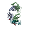
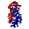
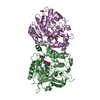
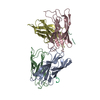
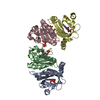
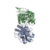
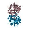
| Title | Crystal structure of beta-lactamase from Sulfitobacter sp. EE-36 | ||||||
 Components Components | beta-lactamase | ||||||
 Keywords Keywords | HYDROLASE / antibiotic resistance / Structural Genomics / Center for Structural Genomics of Infectious Diseases / CSGID | ||||||
| Function / homology |  Function and homology information Function and homology informationantibiotic catabolic process / beta-lactamase activity / beta-lactamase / peptidase activity / outer membrane-bounded periplasmic space / response to antibiotic Similarity search - Function | ||||||
| Biological species |  Sulfitobacter sp. EE-36 (bacteria) Sulfitobacter sp. EE-36 (bacteria) | ||||||
| Method |  X-RAY DIFFRACTION / SYNCHROTRON / SAD / Resolution: 1.55 Å X-RAY DIFFRACTION / SYNCHROTRON / SAD / Resolution: 1.55 Å | ||||||
 Authors Authors | Michalska, K. / Tesar, C. / Endres, M. / Joachimiak, A. / Center for Structural Genomics of Infectious Diseases (CSGID) | ||||||
| Funding support |  United States, 1items United States, 1items
| ||||||
 Citation Citation | Journal: To Be Published Title: Crystal structure of beta-lactamase from Sulfitobacter sp. EE-36 Authors: Michalska, K. / Tesar, C. / Endres, M. / Joachimiak, A. / Center for Structural Genomics of Infectious Diseases (CSGID) | ||||||
| History |
|
- Structure visualization
| Structure viewer | Molecule: MolmilJmol/JSmol |
|---|
- Downloads & links
-Download
| PDBx/mmCIF format | 6njk.cif.gz | 407.7 KB | Display | PDBx/mmCIF format |
|---|---|---|---|---|
| PDB format | pdb6njk.ent.gz | 354.3 KB | Display | PDB format |
| PDBx/mmJSON format | 6njk.json.gz | Tree view | PDBx/mmJSON format | |
| Others |  Other downloads Other downloads |
-Validation report
| Summary document | 6njk_validation.pdf.gz | 437.1 KB | Display | wwPDB validaton report |
|---|---|---|---|---|
| Full document | 6njk_full_validation.pdf.gz | 440.4 KB | Display | |
| Data in XML | 6njk_validation.xml.gz | 30.6 KB | Display | |
| Data in CIF | 6njk_validation.cif.gz | 45.9 KB | Display | |
| Arichive directory | https://data.pdbj.org/pub/pdb/validation_reports/nj/6njkftp://data.pdbj.org/pub/pdb/validation_reports/nj/6njk | HTTPS FTP |
-Related structure data
| Similar structure data | |
|---|---|
| Other databases |


-Links
 PDBj
PDBj
- Assembly
| Deposited unit | 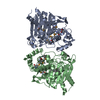
| ||||||||
|---|---|---|---|---|---|---|---|---|---|
| 1 |
| ||||||||
| Unit cell |
|
-Components
| #1: Protein | Mass: 39689.227 Da / Num. of mol.: 2 Source method: isolated from a genetically manipulated source Source: (gene. exp.) Sulfitobacter sp. EE-36 (bacteria) / Gene: EE36_13938 / Plasmid: pMCSG53 / Production host: #2: Chemical | ChemComp-ACT / |   Mass: 59.044 Da / Num. of mol.: 1 / Source method: obtained synthetically / Formula: C2H3O2 Mass: 59.044 Da / Num. of mol.: 1 / Source method: obtained synthetically / Formula: C2H3O2#3: Water | ChemComp-HOH / |  Mass: 18.015 Da / Num. of mol.: 506 / Source method: isolated from a natural source / Formula: H2O Mass: 18.015 Da / Num. of mol.: 506 / Source method: isolated from a natural source / Formula: H2OSequence details | putative beta-lactamase precursor [Sulfitobacter sp. EE-36] GenBank: EAP84172.1 | |
|---|
-Experimental details
-Experiment
| Experiment | Method: X-RAY DIFFRACTION / Number of used crystals: 1 |
|---|
- Sample preparation
| Crystal | Density Matthews: 2.22 Å3/Da / Density % sol: 44.59 % |
|---|---|
| Crystal grow | Temperature: 289 K / Method: vapor diffusion, sitting drop / pH: 8 Details: 0.1 M sodium acetate, 25% PEG4000, 5% 2-propanol, cryo 10% glycerol |
-Data collection
| Diffraction | Mean temperature: 100 K / Serial crystal experiment: N |
|---|---|
| Diffraction source | Source: SYNCHROTRON / Site: APS / Beamline: 19-ID / Wavelength: 0.97918 Å |
| Detector | Type: DECTRIS PILATUS3 X 6M / Detector: PIXEL / Date: Nov 9, 2018 / Details: mirrors |
| Radiation | Monochromator: Si(111) / Protocol: SINGLE WAVELENGTH / Monochromatic (M) / Laue (L): M / Scattering type: x-ray |
| Radiation wavelength | Wavelength: 0.97918 Å / Relative weight: 1 |
| Reflection | Resolution: 1.55→30 Å / Num. obs: 98477 / % possible obs: 98.6 % / Observed criterion σ(I): -3 / Redundancy: 6.6 % / CC1/2: 0.989 / Rmerge(I) obs: 0.106 / Net I/σ(I): 13.7 |
| Reflection shell | Resolution: 1.55→1.58 Å / Redundancy: 5.7 % / Rmerge(I) obs: 1.018 / Mean I/σ(I) obs: 1.65 / Num. unique obs: 4838 / CC1/2: 0.633 / % possible all: 96.9 |
- Processing
| Software |
| |||||||||||||||||||||||||||||||||||||||||||||||||||||||||||||||||||||||||||||||||||||||||||||||||||||||||
|---|---|---|---|---|---|---|---|---|---|---|---|---|---|---|---|---|---|---|---|---|---|---|---|---|---|---|---|---|---|---|---|---|---|---|---|---|---|---|---|---|---|---|---|---|---|---|---|---|---|---|---|---|---|---|---|---|---|---|---|---|---|---|---|---|---|---|---|---|---|---|---|---|---|---|---|---|---|---|---|---|---|---|---|---|---|---|---|---|---|---|---|---|---|---|---|---|---|---|---|---|---|---|---|---|---|---|
| Refinement | Method to determine structure: SAD / Resolution: 1.55→29.948 Å / SU ML: 0.13 / Cross valid method: FREE R-VALUE / σ(F): 1.38 / Phase error: 16.64 / Stereochemistry target values: ML
| |||||||||||||||||||||||||||||||||||||||||||||||||||||||||||||||||||||||||||||||||||||||||||||||||||||||||
| Solvent computation | Shrinkage radii: 0.9 Å / VDW probe radii: 1.11 Å | |||||||||||||||||||||||||||||||||||||||||||||||||||||||||||||||||||||||||||||||||||||||||||||||||||||||||
| Refinement step | Cycle: LAST / Resolution: 1.55→29.948 Å
| |||||||||||||||||||||||||||||||||||||||||||||||||||||||||||||||||||||||||||||||||||||||||||||||||||||||||
| Refine LS restraints |
| |||||||||||||||||||||||||||||||||||||||||||||||||||||||||||||||||||||||||||||||||||||||||||||||||||||||||
| LS refinement shell |
|
