- PDB-2dwx: Co-crystal Structure Analysis of GGA1-GAE with the WNSF motif -
+
Open data
ID or keywords:
Loading...
-
Basic information
Entry
Database: PDB / ID: 2dwx
Title
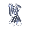
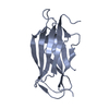
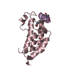
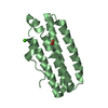
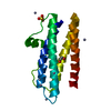
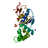
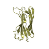
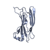
Co-crystal Structure Analysis of GGA1-GAE with the WNSF motif
Components
ADP-ribosylation factor-binding protein GGA1
hinge peptide from ADP-ribosylation factor binding protein GGA1
Keywords
PROTEIN TRANSPORT / IG FOLD / ADAPTIN
Function / homology
Function and homology information
protein localization to ciliary membrane / Golgi to plasma membrane transport / Golgi to plasma membrane protein transport / TBC/RABGAPs / protein localization to cell surface / retrograde transport, endosome to Golgi / phosphatidylinositol binding / protein catabolic process / ubiquitin binding / trans-Golgi network ...protein localization to ciliary membrane / Golgi to plasma membrane transport / Golgi to plasma membrane protein transport / TBC/RABGAPs / protein localization to cell surface / retrograde transport, endosome to Golgi / phosphatidylinositol binding / protein catabolic process / ubiquitin binding / trans-Golgi network / intracellular protein transport / small GTPase binding / positive regulation of protein catabolic process / intracellular protein localization / early endosome membrane / early endosome / endosome membrane / Amyloid fiber formation / Golgi apparatus / protein-containing complex / nucleoplasm / membrane / cytosol Similarity search - Function
ADP-ribosylation factor-binding protein GGA3 / Gamma-adaptin ear (GAE) domain / N-terminal extension of GAT domain / N-terminal extension of GAT domain / GAT domain / GAT domain superfamily / GAT domain / GAT domain profile. / VHS domain / VHS domain ...ADP-ribosylation factor-binding protein GGA3 / Gamma-adaptin ear (GAE) domain / N-terminal extension of GAT domain / N-terminal extension of GAT domain / GAT domain / GAT domain superfamily / GAT domain / GAT domain profile. / VHS domain / VHS domain / VHS domain profile. / Domain present in VPS-27, Hrs and STAM / Gamma-adaptin ear (GAE) domain / Gamma-adaptin ear (GAE) domain profile. / Clathrin adaptor, alpha/beta/gamma-adaptin, appendage, Ig-like subdomain / Adaptin C-terminal domain / Adaptin C-terminal domain / Clathrin adaptor, appendage, Ig-like subdomain superfamily / ENTH/VHS / Immunoglobulin-like / Sandwich / Mainly Beta Similarity search - Domain/homology
#252 - Dec 2020 Hepatitis C Virus Protease/Helicase similarity (1)
-
Assembly
Deposited unit
A: ADP-ribosylation factor-binding protein GGA1 B: ADP-ribosylation factor-binding protein GGA1 C: ADP-ribosylation factor-binding protein GGA1 D: ADP-ribosylation factor-binding protein GGA1 P: hinge peptide from ADP-ribosylation factor binding protein GGA1 Q: hinge peptide from ADP-ribosylation factor binding protein GGA1
In the structure databanks used in Yorodumi, some data are registered as the other names, "COVID-19 virus" and "2019-nCoV". Here are the details of the virus and the list of structure data.
Jan 31, 2019. EMDB accession codes are about to change! (news from PDBe EMDB page)
EMDB accession codes are about to change! (news from PDBe EMDB page)
The allocation of 4 digits for EMDB accession codes will soon come to an end. Whilst these codes will remain in use, new EMDB accession codes will include an additional digit and will expand incrementally as the available range of codes is exhausted. The current 4-digit format prefixed with “EMD-” (i.e. EMD-XXXX) will advance to a 5-digit format (i.e. EMD-XXXXX), and so on. It is currently estimated that the 4-digit codes will be depleted around Spring 2019, at which point the 5-digit format will come into force.
The EM Navigator/Yorodumi systems omit the EMD- prefix.
Related info.:Q: What is EMD? / ID/Accession-code notation in Yorodumi/EM Navigator
Yorodumi is a browser for structure data from EMDB, PDB, SASBDB, etc.
This page is also the successor to EM Navigator detail page, and also detail information page/front-end page for Omokage search.
The word "yorodu" (or yorozu) is an old Japanese word meaning "ten thousand". "mi" (miru) is to see.
Related info.:EMDB / PDB / SASBDB / Comparison of 3 databanks / Yorodumi Search / Aug 31, 2016. New EM Navigator & Yorodumi / Yorodumi Papers / Jmol/JSmol / Function and homology information / Changes in new EM Navigator and Yorodumi
 Movie
Movie Controller


 Open data
 Basic information
 Components
 Keywords
 Function and homology information
 Homo sapiens (human)
 X-RAY DIFFRACTION /
  Authors
 Citation
 Structure visualization
 Downloads & links
 Other downloads


 PDBj
PDBj


 Assembly
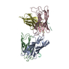


 Mass: 18.015 Da / Num. of mol.: 122 / Source method: isolated from a natural source / Formula: H2O
 Sample preparation
 / Beamline: AR-NW12A / Wavelength: 0.978
  Processing