 Movie
Movie Controller


+ Open data

- Basic information
| Entry | Database: PDB / ID: 1sd9 | ||||||
|---|---|---|---|---|---|---|---|
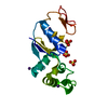
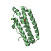
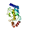
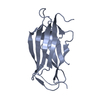
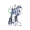
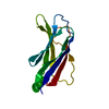
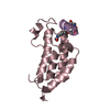
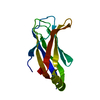
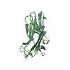
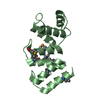
| Title | ARSENATE REDUCTASE C12S MUTANT +0.4M ARSENATE FROM E. COLI | ||||||
 Components Components | Arsenate reductase | ||||||
 Keywords Keywords | OXIDOREDUCTASE / ARSC / REDUCTASE / ARSENITE / ARSENATE | ||||||
| Function / homology |  Function and homology information Function and homology informationarsenate reductase (glutathione/glutaredoxin) / arsenate reductase (glutaredoxin) activity / response to arsenic-containing substance Similarity search - Function | ||||||
| Biological species |  | ||||||
| Method |  X-RAY DIFFRACTION / FOURIER SYNTHESIS / Resolution: 1.65 Å X-RAY DIFFRACTION / FOURIER SYNTHESIS / Resolution: 1.65 Å | ||||||
 Authors Authors | DeMel, S. / Edwards, B.F. | ||||||
 Citation Citation | Journal: Protein Sci. / Year: 2004 Title: Arginine 60 in the ArsC arsenate reductase of E. coli plasmid R773 determines the chemical nature of the bound As(III) product. Authors: DeMel, S. / Shi, J. / Martin, P. / Rosen, B.P. / Edwards, B.F. | ||||||
| History |
|
- Structure visualization
| Structure viewer | Molecule: MolmilJmol/JSmol |
|---|
- Downloads & links
-Download
| PDBx/mmCIF format | 1sd9.cif.gz | 49.3 KB | Display | PDBx/mmCIF format |
|---|---|---|---|---|
| PDB format | pdb1sd9.ent.gz | 34 KB | Display | PDB format |
| PDBx/mmJSON format | 1sd9.json.gz | Tree view | PDBx/mmJSON format | |
| Others |  Other downloads Other downloads |
-Validation report
| Arichive directory | https://data.pdbj.org/pub/pdb/validation_reports/sd/1sd9ftp://data.pdbj.org/pub/pdb/validation_reports/sd/1sd9 | HTTPS FTP |
|---|
-Related structure data
| Related structure data |  1s3cC 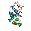 1s3dC 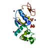 1sd8C 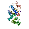 1sjzC 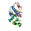 1sk0C 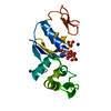 1sk1C 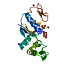 1sk2C C: citing same article ( |
|---|---|
| Similar structure data |
-Links
 PDBj
PDBj- Assembly
| Deposited unit | 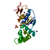
| ||||||||
|---|---|---|---|---|---|---|---|---|---|
| 1 |
| ||||||||
| 2 | 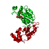
| ||||||||
| Unit cell |
| ||||||||
| Components on special symmetry positions |
|
-Components
| #1: Protein | Mass: 15833.175 Da / Num. of mol.: 1 / Mutation: C12S Source method: isolated from a genetically manipulated source Source: (gene. exp.) Escherichia coli / Production host: References: UniProt: P08692, arsenate reductase (glutathione/glutaredoxin) | ||||
|---|---|---|---|---|---|
| #2: Chemical |   Mass: 96.063 Da / Num. of mol.: 3 / Source method: obtained synthetically / Formula: SO4 Mass: 96.063 Da / Num. of mol.: 3 / Source method: obtained synthetically / Formula: SO4#3: Chemical |   Mass: 132.905 Da / Num. of mol.: 2 / Source method: obtained synthetically / Formula: Cs Mass: 132.905 Da / Num. of mol.: 2 / Source method: obtained synthetically / Formula: Cs#4: Water | ChemComp-HOH / |  Mass: 18.015 Da / Num. of mol.: 305 / Source method: isolated from a natural source / Formula: H2O Mass: 18.015 Da / Num. of mol.: 305 / Source method: isolated from a natural source / Formula: H2O |
-Experimental details
-Experiment
| Experiment | Method: X-RAY DIFFRACTION / Number of used crystals: 1 |
|---|
- Sample preparation
| Crystal | Density Matthews: 2.93 Å3/Da / Density % sol: 58 % |
|---|---|
| Crystal grow | Temperature: 278 K / Method: vapor diffusion, hanging drop / pH: 4.8 Details: 50% saturated Cesium Sulfate, 100mM sodium acetate,5mM DTT, pH 4.80, VAPOR DIFFUSION, HANGING DROP, temperature 278K |
-Data collection
| Diffraction | Mean temperature: 93 K |
|---|---|
| Diffraction source | Source: ROTATING ANODE / Type: RIGAKU RU200 / Wavelength: 1.5418 |
| Detector | Type: RIGAKU RAXIS IV / Detector: IMAGE PLATE / Date: Jan 28, 2002 / Details: OSMIC MIRRORS |
| Radiation | Monochromator: OSMIC MIRRORS / Protocol: SINGLE WAVELENGTH / Monochromatic (M) / Laue (L): M / Scattering type: x-ray |
| Radiation wavelength | Wavelength: 1.5418 Å / Relative weight: 1 |
| Reflection | Resolution: 1.64→20 Å / Num. all: 29169 / Num. obs: 27713 / % possible obs: 90.8 % / Observed criterion σ(I): -3 / Redundancy: 17.9 % / Rmerge(I) obs: 0.166 / Rsym value: 0.166 / Net I/σ(I): 7.7 |
| Reflection shell | Resolution: 1.64→1.698 Å / Redundancy: 6.8 % / Rmerge(I) obs: 0.6117 / Mean I/σ(I) obs: 1.306 / Rsym value: 0.6117 / % possible all: 54.1 |
- Processing
| Software |
| |||||||||||||||||||||||||||||||||
|---|---|---|---|---|---|---|---|---|---|---|---|---|---|---|---|---|---|---|---|---|---|---|---|---|---|---|---|---|---|---|---|---|---|---|
| Refinement | Method to determine structure: FOURIER SYNTHESIS Starting model: NATIVE STRUCTURE Resolution: 1.65→20 Å / Num. parameters: 5908 / Num. restraintsaints: 4595 / Cross valid method: FREE R / σ(I): 0 / Stereochemistry target values: Engh & Huber
| |||||||||||||||||||||||||||||||||
| Solvent computation | Solvent model: MOEWS & KRETSINGER, J.MOL.BIOL.91(1973)201-228 | |||||||||||||||||||||||||||||||||
| Refine analyze | Num. disordered residues: 5 / Occupancy sum hydrogen: 0 / Occupancy sum non hydrogen: 1442.23 | |||||||||||||||||||||||||||||||||
| Refinement step | Cycle: LAST / Resolution: 1.65→20 Å
| |||||||||||||||||||||||||||||||||
| Refine LS restraints |
|
