 Movie
Movie Controller

[English] 日本語
 Yorodumi
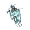
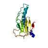
- PDB-5ln4: Crystal structure of self-complemented PsaA, the major subunit of... -
+ Open data

- Basic information
| Entry | Database: PDB / ID: 5ln4 | |||||||||
|---|---|---|---|---|---|---|---|---|---|---|
| Title | Crystal structure of self-complemented PsaA, the major subunit of pH 6 antigen from Yersinia pests, in complex with choline | |||||||||
 Components Components | pH 6 antigen,pH 6 antigen | |||||||||
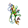
 Keywords Keywords | CELL ADHESION / Ig-like fold / beta sandwich / donor-strand complementation | |||||||||
| Function / homology |  Function and homology information Function and homology information | |||||||||
| Biological species |   Yersinia pestis (bacteria) Yersinia pestis (bacteria) | |||||||||
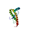
| Method |  X-RAY DIFFRACTION / SYNCHROTRON / MOLECULAR REPLACEMENT / molecular replacement / Resolution: 2.36 Å X-RAY DIFFRACTION / SYNCHROTRON / MOLECULAR REPLACEMENT / molecular replacement / Resolution: 2.36 Å | |||||||||
 Authors Authors | Pakharukova, N.A. / Roy, S. / Rahman, M.M. / Tuitilla, M. / Zavialov, A.V. | |||||||||
| Funding support |  Finland, 2items Finland, 2items
| |||||||||
 Citation Citation | Journal: Mol.Microbiol. / Year: 2016 Title: Structural basis for Myf and Psa fimbriae-mediated tropism of pathogenic strains of Yersinia for host tissues. Authors: Pakharukova, N. / Roy, S. / Tuittila, M. / Rahman, M.M. / Paavilainen, S. / Ingars, A.K. / Skaldin, M. / Lamminmaki, U. / Hard, T. / Teneberg, S. / Zavialov, A.V. | |||||||||
| History |
|
- Structure visualization
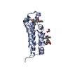
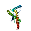
| Structure viewer | Molecule: MolmilJmol/JSmol |
|---|
- Downloads & links
-Download
| PDBx/mmCIF format | 5ln4.cif.gz | 96.3 KB | Display | PDBx/mmCIF format |
|---|---|---|---|---|
| PDB format | pdb5ln4.ent.gz | 73.6 KB | Display | PDB format |
| PDBx/mmJSON format | 5ln4.json.gz | Tree view | PDBx/mmJSON format | |
| Others |  Other downloads Other downloads |
-Validation report
| Arichive directory | https://data.pdbj.org/pub/pdb/validation_reports/ln/5ln4ftp://data.pdbj.org/pub/pdb/validation_reports/ln/5ln4 | HTTPS FTP |
|---|
-Related structure data
| Related structure data |  5ln8C  5lndSC  5lo7C S: Starting model for refinement C: citing same article ( |
|---|---|
| Similar structure data |


-Links
 PDBj
PDBj


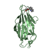
- Assembly
| Deposited unit | 
| ||||||||
|---|---|---|---|---|---|---|---|---|---|
| 1 | 
| ||||||||
| 2 | 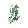
| ||||||||
| 3 | 
| ||||||||
| Unit cell |
|
-Components
| #1: Protein | Mass: 14747.446 Da / Num. of mol.: 3 Source method: isolated from a genetically manipulated source Source: (gene. exp.) Yersinia pestis (bacteria) / Gene: psaA, YPO1303, y2882, YP_1289 / Plasmid: pET101D / Production host: #2: Chemical | 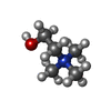  Mass: 104.171 Da / Num. of mol.: 3 / Source method: obtained synthetically / Formula: C5H14NO Mass: 104.171 Da / Num. of mol.: 3 / Source method: obtained synthetically / Formula: C5H14NO#3: Water | ChemComp-HOH / |  Mass: 18.015 Da / Num. of mol.: 185 / Source method: isolated from a natural source / Formula: H2O Mass: 18.015 Da / Num. of mol.: 185 / Source method: isolated from a natural source / Formula: H2O |
|---|
-Experimental details
-Experiment
| Experiment | Method: X-RAY DIFFRACTION / Number of used crystals: 1 |
|---|
- Sample preparation
| Crystal | Density Matthews: 3.56 Å3/Da / Density % sol: 65.45 % / Mosaicity: 0.28 ° |
|---|---|
| Crystal grow | Temperature: 294 K / Method: vapor diffusion, hanging drop / pH: 7.8 Details: 0.1 M imidazole, 0.2 M zinc acetate and 18% PEG3350 |
-Data collection
| Diffraction | Mean temperature: 100 K | ||||||||||||||||||||||||||||||||||||||||||||||||||||||||||||||||||||||||||||||||||||||||||||||||
|---|---|---|---|---|---|---|---|---|---|---|---|---|---|---|---|---|---|---|---|---|---|---|---|---|---|---|---|---|---|---|---|---|---|---|---|---|---|---|---|---|---|---|---|---|---|---|---|---|---|---|---|---|---|---|---|---|---|---|---|---|---|---|---|---|---|---|---|---|---|---|---|---|---|---|---|---|---|---|---|---|---|---|---|---|---|---|---|---|---|---|---|---|---|---|---|---|---|
| Diffraction source | Source: SYNCHROTRON / Site: ESRF  / Beamline: ID29 / Wavelength: 0.976 Å / Beamline: ID29 / Wavelength: 0.976 Å | ||||||||||||||||||||||||||||||||||||||||||||||||||||||||||||||||||||||||||||||||||||||||||||||||
| Detector | Type: DECTRIS PILATUS3 6M / Detector: PIXEL / Date: Dec 7, 2012 | ||||||||||||||||||||||||||||||||||||||||||||||||||||||||||||||||||||||||||||||||||||||||||||||||
| Radiation | Protocol: SINGLE WAVELENGTH / Monochromatic (M) / Laue (L): M / Scattering type: x-ray | ||||||||||||||||||||||||||||||||||||||||||||||||||||||||||||||||||||||||||||||||||||||||||||||||
| Radiation wavelength | Wavelength: 0.976 Å / Relative weight: 1 | ||||||||||||||||||||||||||||||||||||||||||||||||||||||||||||||||||||||||||||||||||||||||||||||||
| Reflection | Resolution: 2.36→49.4 Å / Num. obs: 25917 / % possible obs: 99.5 % / Redundancy: 3.5 % / Rsym value: 0.082 / Net I/av σ(I): 8.857 / Net I/σ(I): 11.2 | ||||||||||||||||||||||||||||||||||||||||||||||||||||||||||||||||||||||||||||||||||||||||||||||||
| Reflection shell |
|
-Phasing
| Phasing | Method: molecular replacement |
|---|
- Processing
| Software |
| ||||||||||||||||||||||||||||||||||||||||||||||||||||||||||||
|---|---|---|---|---|---|---|---|---|---|---|---|---|---|---|---|---|---|---|---|---|---|---|---|---|---|---|---|---|---|---|---|---|---|---|---|---|---|---|---|---|---|---|---|---|---|---|---|---|---|---|---|---|---|---|---|---|---|---|---|---|---|
| Refinement | Method to determine structure: MOLECULAR REPLACEMENT Starting model: 5LND Resolution: 2.36→49.397 Å / Cor.coef. Fo:Fc: 0.924 / Cor.coef. Fo:Fc free: 0.906 / SU B: 8.276 / SU ML: 0.191 / Cross valid method: THROUGHOUT / σ(F): 0 / ESU R: 0.299 / ESU R Free: 0.237 / Details: U VALUES : REFINED INDIVIDUALLY
| ||||||||||||||||||||||||||||||||||||||||||||||||||||||||||||
| Solvent computation | Ion probe radii: 0.8 Å / Shrinkage radii: 0.8 Å / VDW probe radii: 1.2 Å | ||||||||||||||||||||||||||||||||||||||||||||||||||||||||||||
| Displacement parameters | Biso max: 147.92 Å2 / Biso mean: 44.315 Å2 / Biso min: 8.12 Å2
| ||||||||||||||||||||||||||||||||||||||||||||||||||||||||||||
| Refinement step | Cycle: final / Resolution: 2.36→49.397 Å
| ||||||||||||||||||||||||||||||||||||||||||||||||||||||||||||
| Refine LS restraints |
| ||||||||||||||||||||||||||||||||||||||||||||||||||||||||||||
| LS refinement shell | Resolution: 2.36→2.421 Å / Total num. of bins used: 20
|
