Type: MARMOSAIC 300 mm CCD / Detector: CCD / Date: Nov 1, 2010
Radiation
Protocol: SINGLE WAVELENGTH / Monochromatic (M) / Laue (L): M / Scattering type: x-ray
Radiation wavelength
Wavelength: 1 Å / Relative weight: 1
Reflection
Redundancy: 2.6 % / Av σ(I) over netI: 6.29 / Number: 41798 / Rmerge(I) obs: 0.115 / Χ2: 0.97 / D res high: 1.61 Å / D res low: 50 Å / Num. obs: 16080 / % possible obs: 92.5
Diffraction reflection shell
Highest resolution (Å)
Lowest resolution (Å)
% possible obs (%)
ID
Rmerge(I) obs
Chi squared
Redundancy
3.47
50
95.7
1
0.054
0.953
3.2
2.75
3.47
96.1
1
0.085
0.934
3
2.4
2.75
96.4
1
0.151
1.01
3
2.19
2.4
95.9
1
0.212
1.041
2.9
2.03
2.19
93.5
1
0.27
1.042
2.8
1.91
2.03
93.9
1
0.34
1.075
2.5
1.81
1.91
91.6
1
0.394
1.092
2.3
1.73
1.81
89.5
1
0.432
1.002
2.1
1.67
1.73
86.4
1
0.453
0.725
2
1.61
1.67
84.7
1
0.467
0.616
1.9
Reflection
Resolution: 1.61→50 Å / Num. obs: 16080 / % possible obs: 92.5 % / Redundancy: 2.6 % / Rmerge(I) obs: 0.115 / Χ2: 0.974 / Net I/σ(I): 7.4
Reflection shell
Resolution (Å)
Redundancy (%)
Rmerge(I) obs
Num. unique all
Χ2
Diffraction-ID
% possible all
1.61-1.67
1.9
0.467
1465
0.616
1
84.7
1.67-1.73
2
0.453
1432
0.725
1
86.4
1.73-1.81
2.1
0.432
1527
1.002
1
89.5
1.81-1.91
2.3
0.394
1577
1.092
1
91.6
1.91-2.03
2.5
0.34
1589
1.075
1
93.9
2.03-2.19
2.8
0.27
1607
1.042
1
93.5
2.19-2.4
2.9
0.212
1664
1.041
1
95.9
2.4-2.75
3
0.151
1703
1.01
1
96.4
2.75-3.47
3
0.085
1694
0.934
1
96.1
3.47-50
3.2
0.054
1822
0.953
1
95.7
-
Phasing
Phasing
Method: molecular replacement
-
Processing
Software
Name
Version
Classification
NB
DENZO
datareduction
SCALEPACK
datascaling
PHASER
phasing
REFMAC
refinement
PDB_EXTRACT
3.11
dataextraction
HKL-2000
datacollection
PHENIX
1.7.2_869
refinement
Refinement
Method to determine structure: MOLECULAR REPLACEMENT / Resolution: 1.9→15.387 Å / Occupancy max: 1 / Occupancy min: 0.32 / SU ML: 0.69 / σ(F): 1.46 / Phase error: 26.91 / Stereochemistry target values: ML
Rfactor
Num. reflection
% reflection
Rfree
0.2863
598
4.98 %
Rwork
0.2411
-
-
obs
0.2433
12006
94.99 %
Solvent computation
Shrinkage radii: 0.73 Å / VDW probe radii: 1 Å / Solvent model: FLAT BULK SOLVENT MODEL / Bsol: 44.659 Å2 / ksol: 0.377 e/Å3
In the structure databanks used in Yorodumi, some data are registered as the other names, "COVID-19 virus" and "2019-nCoV". Here are the details of the virus and the list of structure data.
Jan 31, 2019. EMDB accession codes are about to change! (news from PDBe EMDB page)
EMDB accession codes are about to change! (news from PDBe EMDB page)
The allocation of 4 digits for EMDB accession codes will soon come to an end. Whilst these codes will remain in use, new EMDB accession codes will include an additional digit and will expand incrementally as the available range of codes is exhausted. The current 4-digit format prefixed with “EMD-” (i.e. EMD-XXXX) will advance to a 5-digit format (i.e. EMD-XXXXX), and so on. It is currently estimated that the 4-digit codes will be depleted around Spring 2019, at which point the 5-digit format will come into force.
The EM Navigator/Yorodumi systems omit the EMD- prefix.
Related info.:Q: What is EMD? / ID/Accession-code notation in Yorodumi/EM Navigator
Yorodumi is a browser for structure data from EMDB, PDB, SASBDB, etc.
This page is also the successor to EM Navigator detail page, and also detail information page/front-end page for Omokage search.
The word "yorodu" (or yorozu) is an old Japanese word meaning "ten thousand". "mi" (miru) is to see.
Related info.:EMDB / PDB / SASBDB / Comparison of 3 databanks / Yorodumi Search / Aug 31, 2016. New EM Navigator & Yorodumi / Yorodumi Papers / Jmol/JSmol / Function and homology information / Changes in new EM Navigator and Yorodumi
 Movie
Movie Controller

 Yorodumi
 Open data
 Basic information
 Components
 Keywords
 Function and homology information
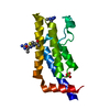
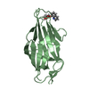
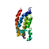
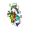
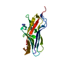
 Yersinia pestis (bacteria)
 X-RAY DIFFRACTION /
  Authors
 Citation
 Structure visualization
 Downloads & links
 Other downloads


 PDBj
PDBj


 Assembly
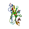


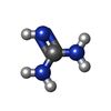


 Mass: 59.070 Da / Num. of mol.: 2 / Source method: obtained synthetically / Formula: CH5N3
 Mass: 35.453 Da / Num. of mol.: 1 / Source method: obtained synthetically / Formula: Cl
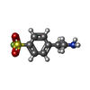
 Mass: 203.234 Da / Num. of mol.: 1 / Source method: obtained synthetically / Formula: C8H10FNO2S / Comment: protease inhibitor*YM
 Sample preparation
 / Beamline: 22-ID / Wavelength: 1 Å
 Processing