 Movie
Movie Controller

[English] 日本語
 Yorodumi
- PDB-6mtg: A Single Reactive Noncanonical Amino Acid is Able to Dramatically... -
+ Open data

- Basic information
| Entry | Database: PDB / ID: 6mtg | ||||||
|---|---|---|---|---|---|---|---|
| Title | A Single Reactive Noncanonical Amino Acid is Able to Dramatically Stabilize Protein Structure | ||||||
 Components Components | Homoserine O-succinyltransferase | ||||||
 Keywords Keywords | TRANSFERASE / noncanonical amino acid / isothiocyanate / crosslink / thiourea / stabilization | ||||||
| Function / homology |  Function and homology informationhomoserine O-succinyltransferase / homoserine O-succinyltransferase activity / L-methionine biosynthetic process from homoserine via O-succinyl-L-homoserine and cystathionine / homoserine O-acetyltransferase activity / cytoplasm Function and homology informationhomoserine O-succinyltransferase / homoserine O-succinyltransferase activity / L-methionine biosynthetic process from homoserine via O-succinyl-L-homoserine and cystathionine / homoserine O-acetyltransferase activity / cytoplasmSimilarity search - Function | ||||||
| Biological species |  Escherichia coli (E. coli) Escherichia coli (E. coli) | ||||||
| Method | X-RAY DIFFRACTION / SYNCHROTRON / MOLECULAR REPLACEMENT / molecular replacement / Resolution: 1.85 Å | ||||||
 Authors Authors | Li, J.C. / Nasertorabi, F. / Xuan, W. / Han, G.W. / Stevens, R.C. / Schultz, P.G. | ||||||
| Funding support |  United States, 1items United States, 1items
| ||||||
 Citation Citation | Journal: Acs Chem.Biol. / Year: 2019 Title: A Single Reactive Noncanonical Amino Acid Is Able to Dramatically Stabilize Protein Structure. Authors: Li, J.C. / Nastertorabi, F. / Xuan, W. / Han, G.W. / Stevens, R.C. / Schultz, P.G. | ||||||
| History |
|
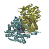
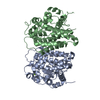
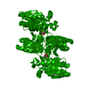
- Structure visualization
| Structure viewer | Molecule: MolmilJmol/JSmol |
|---|
- Downloads & links
-Download
| PDBx/mmCIF format | 6mtg.cif.gz | 257 KB | Display | PDBx/mmCIF format |
|---|---|---|---|---|
| PDB format | pdb6mtg.ent.gz | 205 KB | Display | PDB format |
| PDBx/mmJSON format | 6mtg.json.gz | Tree view | PDBx/mmJSON format | |
| Others |  Other downloads Other downloads |
-Validation report
| Arichive directory | https://data.pdbj.org/pub/pdb/validation_reports/mt/6mtgftp://data.pdbj.org/pub/pdb/validation_reports/mt/6mtg | HTTPS FTP |
|---|
-Related structure data
| Related structure data | 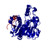 2h2wS S: Starting model for refinement |
|---|---|
| Similar structure data |

-Links
 PDBj
PDBj

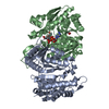
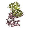
- Assembly
| Deposited unit | 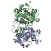
| ||||||||||||||||||
|---|---|---|---|---|---|---|---|---|---|---|---|---|---|---|---|---|---|---|---|
| 1 |
| ||||||||||||||||||
| Unit cell |
| ||||||||||||||||||
| Noncrystallographic symmetry (NCS) | NCS domain:
NCS domain segments: Component-ID: 0 / Ens-ID: 1 / Beg auth comp-ID: PRO / Beg label comp-ID: PRO / End auth comp-ID: ILE / End label comp-ID: ILE / Refine code: 0 / Auth seq-ID: 2 - 296 / Label seq-ID: 2 - 296
|
-Components
| #1: Protein | / HST / Homoserine transsuccinylase / HTS Mass: 35084.328 Da / Num. of mol.: 2 / Mutation: P257I Source method: isolated from a genetically manipulated source Source: (gene. exp.) Escherichia coli (strain K12) (bacteria)Strain: K12 / Gene: metAS, metA, b4013, JW3973 / Plasmid: Modified PET22b / Details (production host): Inserted a T5 promotor / Production host: Escherichia coli K-12 (bacteria)References: UniProt: P07623, homoserine O-succinyltransferase#2: Chemical | Diethylene glycol  Mass: 106.120 Da / Num. of mol.: 2 / Source method: obtained synthetically / Formula: C4H10O3 Mass: 106.120 Da / Num. of mol.: 2 / Source method: obtained synthetically / Formula: C4H10O3#3: Chemical | ChemComp-FMT / Formic acid  Mass: 46.025 Da / Num. of mol.: 5 / Source method: obtained synthetically / Formula: CH2O2 Mass: 46.025 Da / Num. of mol.: 5 / Source method: obtained synthetically / Formula: CH2O2#4: Chemical | ChemComp-GOL / | Glycerol  Mass: 92.094 Da / Num. of mol.: 1 Mass: 92.094 Da / Num. of mol.: 1Source method: isolated from a genetically manipulated source Formula: C3H8O3 #5: Water | ChemComp-HOH / | Water Mass: 18.015 Da / Num. of mol.: 544 / Source method: isolated from a natural source / Formula: H2O Mass: 18.015 Da / Num. of mol.: 544 / Source method: isolated from a natural source / Formula: H2O |
|---|
-Experimental details
-Experiment
| Experiment | Method: X-RAY DIFFRACTION / Number of used crystals: 1 |
|---|
- Sample preparation
| Crystal | Density Matthews: 2.35 Å3/Da / Density % sol: 47.57 % / Description: Thin Hexagonal |
|---|---|
| Crystal grow | Temperature: 277 K / Method: vapor diffusion, sitting drop / pH: 7.4 / Details: 100mM Potassium Formate and 26% PEG 3350 / PH range: 7.2-7.8 |
-Data collection
| Diffraction | Mean temperature: 100 K / Ambient temp details: Cryo / Serial crystal experiment: N | ||||||||||||||||||||||||||||||||||||||||||||||||||||||||||||||||||||||||||||||||||||||||||||||||||||||||||||||
|---|---|---|---|---|---|---|---|---|---|---|---|---|---|---|---|---|---|---|---|---|---|---|---|---|---|---|---|---|---|---|---|---|---|---|---|---|---|---|---|---|---|---|---|---|---|---|---|---|---|---|---|---|---|---|---|---|---|---|---|---|---|---|---|---|---|---|---|---|---|---|---|---|---|---|---|---|---|---|---|---|---|---|---|---|---|---|---|---|---|---|---|---|---|---|---|---|---|---|---|---|---|---|---|---|---|---|---|---|---|---|---|
| Diffraction source | Source: SYNCHROTRON / Site: SSRL / Beamline: BL12-2 / Wavelength: 0.9795 Å | ||||||||||||||||||||||||||||||||||||||||||||||||||||||||||||||||||||||||||||||||||||||||||||||||||||||||||||||
| Detector | Type: DECTRIS PILATUS 6M / Detector: PIXEL / Date: Apr 29, 2018 Details: Flat Si Rh coated M0, Kirkpatrick-Baez flat bent Si M1 & M2 | ||||||||||||||||||||||||||||||||||||||||||||||||||||||||||||||||||||||||||||||||||||||||||||||||||||||||||||||
| Radiation | Monochromator: Liquid nitrogen-cooled double crystal Si(111) Protocol: SINGLE WAVELENGTH / Monochromatic (M) / Laue (L): M / Scattering type: x-ray | ||||||||||||||||||||||||||||||||||||||||||||||||||||||||||||||||||||||||||||||||||||||||||||||||||||||||||||||
| Radiation wavelength | Wavelength: 0.9795 Å / Relative weight: 1 | ||||||||||||||||||||||||||||||||||||||||||||||||||||||||||||||||||||||||||||||||||||||||||||||||||||||||||||||
| Reflection | Resolution: 1.85→78.157 Å / Num. obs: 56066 / % possible obs: 99.3 % / Redundancy: 13.4 % / CC1/2: 0.998 / Rpim(I) all: 0.044 / Rrim(I) all: 0.162 / Rsym value: 0.156 / Net I/av σ(I): 4.1 / Net I/σ(I): 11.5 | ||||||||||||||||||||||||||||||||||||||||||||||||||||||||||||||||||||||||||||||||||||||||||||||||||||||||||||||
| Reflection shell | Diffraction-ID: 1
|
-Phasing
| Phasing | Method: molecular replacement |
|---|
- Processing
| Software |
| ||||||||||||||||||||||||||||||||||||||||||||||||||||||||||||||||||||||||
|---|---|---|---|---|---|---|---|---|---|---|---|---|---|---|---|---|---|---|---|---|---|---|---|---|---|---|---|---|---|---|---|---|---|---|---|---|---|---|---|---|---|---|---|---|---|---|---|---|---|---|---|---|---|---|---|---|---|---|---|---|---|---|---|---|---|---|---|---|---|---|---|---|---|
| Refinement | Method to determine structure: MOLECULAR REPLACEMENT Starting model: 2H2W Resolution: 1.85→29.89 Å / Cor.coef. Fo:Fc: 0.966 / Cor.coef. Fo:Fc free: 0.945 / SU B: 6.819 / SU ML: 0.102 / Cross valid method: THROUGHOUT / σ(F): 0 / ESU R: 0.132 / ESU R Free: 0.131 Details: HYDROGENS HAVE BEEN ADDED IN THE RIDING POSITIONS U VALUES : WITH TLS ADDED
| ||||||||||||||||||||||||||||||||||||||||||||||||||||||||||||||||||||||||
| Solvent computation | Ion probe radii: 0.8 Å / Shrinkage radii: 0.8 Å / VDW probe radii: 1.2 Å | ||||||||||||||||||||||||||||||||||||||||||||||||||||||||||||||||||||||||
| Displacement parameters | Biso max: 79.86 Å2 / Biso mean: 28.1 Å2 / Biso min: 16.82 Å2
| ||||||||||||||||||||||||||||||||||||||||||||||||||||||||||||||||||||||||
| Refinement step | Cycle: final / Resolution: 1.85→29.89 Å
| ||||||||||||||||||||||||||||||||||||||||||||||||||||||||||||||||||||||||
| Refine LS restraints |
| ||||||||||||||||||||||||||||||||||||||||||||||||||||||||||||||||||||||||
| Refine LS restraints NCS | Ens-ID: 1 / Number: 9086 / Refine-ID: X-RAY DIFFRACTION / Type: interatomic distance / Rms dev position: 0.12 Å / Weight position: 0.05
| ||||||||||||||||||||||||||||||||||||||||||||||||||||||||||||||||||||||||
| LS refinement shell | Resolution: 1.85→1.898 Å / Rfactor Rfree error: 0 / Total num. of bins used: 20
| ||||||||||||||||||||||||||||||||||||||||||||||||||||||||||||||||||||||||
| Refinement TLS params. | L12: -0.0241 °2 / Method: refined / Refine-ID: X-RAY DIFFRACTION
| ||||||||||||||||||||||||||||||||||||||||||||||||||||||||||||||||||||||||
| Refinement TLS group |
|
