 Movie
Movie Controller

[English] 日本語
 Yorodumi
- PDB-2h2w: Crystal structure of Homoserine O-succinyltransferase (EC 2.3.1.4... -
+ Open data

- Basic information
| Entry | Database: PDB / ID: 2h2w | ||||||
|---|---|---|---|---|---|---|---|
| Title | Crystal structure of Homoserine O-succinyltransferase (EC 2.3.1.46) (Homoserine O-transsuccinylase) (HTS) (tm0881) from THERMOTOGA MARITIMA at 2.52 A resolution | ||||||
 Components Components | Homoserine O-succinyltransferase | ||||||
 Keywords Keywords | TRANSFERASE / tm0881 / Homoserine O-succinyltransferase / (EC 2.3.1.46) / Homoserine O-transsuccinylase / HTS / (tm0881) / Structural Genomics / Joint Center for Structural Genomics / JCSG / Protein Structure Initiative / PSI | ||||||
| Function / homology |  Function and homology information Function and homology informationhomoserine O-succinyltransferase activity / : / homoserine O-acetyltransferase / homoserine O-acetyltransferase activity / cytoplasm Similarity search - Function | ||||||
| Biological species |   Thermotoga maritima (bacteria) Thermotoga maritima (bacteria) | ||||||
| Method |  X-RAY DIFFRACTION / SYNCHROTRON / MOLECULAR REPLACEMENT / molecular replacement / Resolution: 2.52 Å X-RAY DIFFRACTION / SYNCHROTRON / MOLECULAR REPLACEMENT / molecular replacement / Resolution: 2.52 Å | ||||||
 Authors Authors | Joint Center for Structural Genomics (JCSG) | ||||||
 Citation Citation | Journal: To be published Title: Crystal structure of Homoserine O-succinyltransferase (EC 2.3.1.46) (Homoserine O-transsuccinylase) (HTS) (tm0881) from THERMOTOGA MARITIMA at 2.52 A resolution Authors: Joint Center for Structural Genomics (JCSG) | ||||||
| History |
| ||||||
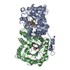
| Remark 300 | BIOMOLECULE: 1 THIS ENTRY CONTAINS THE CRYSTALLOGRAPHIC ASYMMETRIC UNIT WHICH CONSISTS OF 1 CHAIN(S) ...BIOMOLECULE: 1 THIS ENTRY CONTAINS THE CRYSTALLOGRAPHIC ASYMMETRIC UNIT WHICH CONSISTS OF 1 CHAIN(S). SEE REMARK 350 FOR INFORMATION ON GENERATING THE BIOLOGICAL MOLECULE(S). SIZE EXCLUSION CHROMATOGRAPHY WITH STATIC LIGHT SCATTERING SUPPORTS THE ASSIGNMENT OF A DIMER AS A BIOLOGICALLY SIGNIFICANT OLIGIMERIZATION STATE. | ||||||
| Remark 999 | SEQUENCE THE CONSTRUCT WAS EXPRESSED WITH A PURIFICATION TAG MGSDKIHHHHHH FOLLOWED BY THE RESIDUES ...SEQUENCE THE CONSTRUCT WAS EXPRESSED WITH A PURIFICATION TAG MGSDKIHHHHHH FOLLOWED BY THE RESIDUES 1-300 OF THE TARGET SEQUENCE. THE CONSTRUCT WITH RESIDUES 301-304 ELIMINATED PRODUCED THE BEST DIFFRACTING CRYSTAL. |
- Structure visualization
| Structure viewer | Molecule: MolmilJmol/JSmol |
|---|
- Downloads & links
-Download
| PDBx/mmCIF format | 2h2w.cif.gz | 74.1 KB | Display | PDBx/mmCIF format |
|---|---|---|---|---|
| PDB format | pdb2h2w.ent.gz | 53.6 KB | Display | PDB format |
| PDBx/mmJSON format | 2h2w.json.gz | Tree view | PDBx/mmJSON format | |
| Others |  Other downloads Other downloads |
-Validation report
| Arichive directory | https://data.pdbj.org/pub/pdb/validation_reports/h2/2h2wftp://data.pdbj.org/pub/pdb/validation_reports/h2/2h2w | HTTPS FTP |
|---|
-Related structure data
| Related structure data | 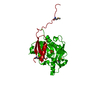 2ghrS S: Starting model for refinement |
|---|---|
| Similar structure data | |
| Other databases |


-Links
 PDBj
PDBj

- Assembly
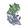
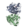
| Deposited unit | 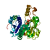
| ||||||||
|---|---|---|---|---|---|---|---|---|---|
| 1 | 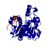
| ||||||||
| Unit cell |
| ||||||||
| Details | SIZE EXCLUSION CHROMATOGRAPHY WITH STATIC LIGHT SCATTERING SUPPORTS THE ASSIGNMENT OF A DIMER AS A BIOLOGICALLY SIGNIFICANT OLIGIMERIZATION STATE. |
-Components
| #1: Protein | Mass: 36772.133 Da / Num. of mol.: 1 Source method: isolated from a genetically manipulated source Source: (gene. exp.) Thermotoga maritima (bacteria) / Gene: metA / Production host: References: UniProt: Q9WZY3, homoserine O-succinyltransferase |
|---|---|
| #2: Water | ChemComp-HOH /  Mass: 18.015 Da / Num. of mol.: 16 / Source method: isolated from a natural source / Formula: H2O Mass: 18.015 Da / Num. of mol.: 16 / Source method: isolated from a natural source / Formula: H2O |
-Experimental details
-Experiment
| Experiment | Method: X-RAY DIFFRACTION / Number of used crystals: 1 |
|---|
- Sample preparation
| Crystal | Density Matthews: 2.88 Å3/Da / Density % sol: 56.96 % |
|---|---|
| Crystal grow | Temperature: 293 K / Method: vapor diffusion, sitting drop, nanodrop / pH: 8 Details: 10.0% iso-Propanol, 0.1M Imidazole pH 8.0, VAPOR DIFFUSION,SITTING DROP,NANODROP, temperature 293K |
-Data collection
| Diffraction | Mean temperature: 100 K | |||||||||||||||||||||||||||||||||||||||||||||||||||||||||||||||||||||||||||||
|---|---|---|---|---|---|---|---|---|---|---|---|---|---|---|---|---|---|---|---|---|---|---|---|---|---|---|---|---|---|---|---|---|---|---|---|---|---|---|---|---|---|---|---|---|---|---|---|---|---|---|---|---|---|---|---|---|---|---|---|---|---|---|---|---|---|---|---|---|---|---|---|---|---|---|---|---|---|---|
| Diffraction source | Source: SYNCHROTRON / Site: ALS  / Beamline: 8.2.1 / Wavelength: 1 Å / Beamline: 8.2.1 / Wavelength: 1 Å | |||||||||||||||||||||||||||||||||||||||||||||||||||||||||||||||||||||||||||||
| Detector | Type: ADSC QUANTUM 210 / Detector: CCD / Date: Sep 21, 2003 | |||||||||||||||||||||||||||||||||||||||||||||||||||||||||||||||||||||||||||||
| Radiation | Monochromator: Double Crystal Si(111) / Protocol: SINGLE WAVELENGTH / Monochromatic (M) / Laue (L): M / Scattering type: x-ray | |||||||||||||||||||||||||||||||||||||||||||||||||||||||||||||||||||||||||||||
| Radiation wavelength | Wavelength: 1 Å / Relative weight: 1 | |||||||||||||||||||||||||||||||||||||||||||||||||||||||||||||||||||||||||||||
| Reflection | Resolution: 2.52→42.3 Å / Num. obs: 13643 / % possible obs: 99.9 % / Redundancy: 10.98 % / Biso Wilson estimate: 70.22 Å2 / Rmerge(I) obs: 0.046 / Χ2: 1.481 / Net I/σ(I): 18.5 | |||||||||||||||||||||||||||||||||||||||||||||||||||||||||||||||||||||||||||||
| Reflection shell | Diffraction-ID: 1
|
-Phasing
| Phasing | Method: molecular replacement |
|---|
- Processing
| Software |
| |||||||||||||||||||||||||||||||||||||||||||||||||||||||||||||||||||||||||||||||||||||||||||||||||||||||||||||||||||||||||||||
|---|---|---|---|---|---|---|---|---|---|---|---|---|---|---|---|---|---|---|---|---|---|---|---|---|---|---|---|---|---|---|---|---|---|---|---|---|---|---|---|---|---|---|---|---|---|---|---|---|---|---|---|---|---|---|---|---|---|---|---|---|---|---|---|---|---|---|---|---|---|---|---|---|---|---|---|---|---|---|---|---|---|---|---|---|---|---|---|---|---|---|---|---|---|---|---|---|---|---|---|---|---|---|---|---|---|---|---|---|---|---|---|---|---|---|---|---|---|---|---|---|---|---|---|---|---|---|
| Refinement | Method to determine structure: MOLECULAR REPLACEMENT Starting model: 2GHR Resolution: 2.52→42.3 Å / Cor.coef. Fo:Fc: 0.963 / Cor.coef. Fo:Fc free: 0.915 / SU B: 21.895 / SU ML: 0.221 / TLS residual ADP flag: LIKELY RESIDUAL / Cross valid method: THROUGHOUT / σ(F): 0 / ESU R: 0.502 / ESU R Free: 0.322 / Stereochemistry target values: MAXIMUM LIKELIHOOD Details: 1. HYDROGENS HAVE BEEN ADDED IN THE RIDING POSITIONS 2. DUE TO STRONG SALT AND SOLVENT RINGS, 1024 REFLECTIONS BETWEEN 2.90-3.02 ANGSTROMS WERE OMITTED FROM THE REFINEMENT. 3. THE LARGE GAP ...Details: 1. HYDROGENS HAVE BEEN ADDED IN THE RIDING POSITIONS 2. DUE TO STRONG SALT AND SOLVENT RINGS, 1024 REFLECTIONS BETWEEN 2.90-3.02 ANGSTROMS WERE OMITTED FROM THE REFINEMENT. 3. THE LARGE GAP BETWEEN THE RWORK AND RFREE IS LIKELY DUE TO ARTIFACTS IN THE DIFFRACTION IMAGES. 4. ATOM RECORDS CONTAIN RESIDUAL B FACTORS ONLY.
| |||||||||||||||||||||||||||||||||||||||||||||||||||||||||||||||||||||||||||||||||||||||||||||||||||||||||||||||||||||||||||||
| Solvent computation | Ion probe radii: 0.8 Å / Shrinkage radii: 0.8 Å / VDW probe radii: 1.2 Å / Solvent model: MASK | |||||||||||||||||||||||||||||||||||||||||||||||||||||||||||||||||||||||||||||||||||||||||||||||||||||||||||||||||||||||||||||
| Displacement parameters | Biso mean: 44.358 Å2
| |||||||||||||||||||||||||||||||||||||||||||||||||||||||||||||||||||||||||||||||||||||||||||||||||||||||||||||||||||||||||||||
| Refinement step | Cycle: LAST / Resolution: 2.52→42.3 Å
| |||||||||||||||||||||||||||||||||||||||||||||||||||||||||||||||||||||||||||||||||||||||||||||||||||||||||||||||||||||||||||||
| Refine LS restraints |
| |||||||||||||||||||||||||||||||||||||||||||||||||||||||||||||||||||||||||||||||||||||||||||||||||||||||||||||||||||||||||||||
| LS refinement shell | Resolution: 2.52→2.584 Å / Total num. of bins used: 20
| |||||||||||||||||||||||||||||||||||||||||||||||||||||||||||||||||||||||||||||||||||||||||||||||||||||||||||||||||||||||||||||
| Refinement TLS params. | Method: refined / Refine-ID: X-RAY DIFFRACTION
| |||||||||||||||||||||||||||||||||||||||||||||||||||||||||||||||||||||||||||||||||||||||||||||||||||||||||||||||||||||||||||||
| Refinement TLS group | Refine-ID: X-RAY DIFFRACTION / Selection: ALL / Auth asym-ID: A / Label asym-ID: A
|
