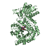
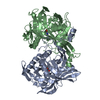
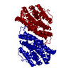
MetA family / Homoserine O-succinyltransferase / Class I glutamine amidotransferase (GATase) domain / Class I glutamine amidotransferase-like / Rossmann fold / 3-Layer(aba) Sandwich / Alpha Beta Similarity search - Domain/homology
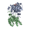
BIOMOLECULE: 1 THIS ENTRY CONTAINS THE CRYSTALLOGRAPHIC ASYMMETRIC UNIT WHICH CONSISTS OF 1 CHAIN(S) ...BIOMOLECULE: 1 THIS ENTRY CONTAINS THE CRYSTALLOGRAPHIC ASYMMETRIC UNIT WHICH CONSISTS OF 1 CHAIN(S). SEE REMARK 350 FOR INFORMATION ON GENERATING THE BIOLOGICAL MOLECULE(S). SIZE EXCLUSION CHROMATOGRAPHY WITH STATIC LIGHT SCATTERING SUPPORTS THE ASSIGNMENT OF A DIMER AS A BIOLOGICALLY SIGNIFICANT OLIGIMERIZATION STATE.
Remark 999
SEQUENCE THE CONSTRUCT WAS EXPRESSED WITH A PURIFICATION TAG MGSDKIHHHHHHENLYFQG. THE TAG WAS ...SEQUENCE THE CONSTRUCT WAS EXPRESSED WITH A PURIFICATION TAG MGSDKIHHHHHHENLYFQG. THE TAG WAS REMOVED WITH TEV PROTEASE LEAVING ONLY A GLYCINE (0) FOLLOWED BY THE TARGET SEQUENCE.
Resolution: 2.4→29.64 Å / Num. obs: 14274 / % possible obs: 99.8 % / Redundancy: 6.6 % / Biso Wilson estimate: 38.1 Å2 / Rmerge(I) obs: 0.124 / Rsym value: 0.124 / Net I/σ(I): 4.6
Reflection shell
Diffraction-ID: 1
Resolution (Å)
% possible obs (%)
Redundancy (%)
Rmerge(I) obs
Mean I/σ(I) obs
Num. measured obs
Rsym value
2.4-2.46
100
6.8
0.592
1.3
1038
0.592
2.46-2.53
100
6.8
0.532
1.4
1001
0.532
2.53-2.6
100
6.8
0.523
1.5
989
0.523
2.6-2.68
100
6.9
0.431
1.8
942
0.431
2.68-2.77
100
6.9
0.36
2.1
924
0.36
2.77-2.87
100
6.8
0.321
2.4
890
0.321
2.87-2.98
100
6.8
0.27
2.8
889
0.27
2.98-3.1
100
6.8
0.187
4.1
829
0.187
3.1-3.24
100
6.8
0.155
4.9
806
0.155
3.24-3.39
100
6.8
0.122
6.3
778
0.122
3.39-3.58
100
6.7
0.096
7.7
729
0.096
3.58-3.79
99.3
5.7
0.135
3.7
695
0.135
3.79-4.06
99.8
6.5
0.075
9.1
662
0.075
4.06-4.38
100
6.5
0.054
12.8
621
0.054
4.38-4.8
99.9
6.4
0.047
13.8
568
0.047
4.8-5.37
99.8
6.2
0.051
13
526
0.051
5.37-6.2
99
5.5
0.068
8.9
464
0.068
6.2-7.59
99.7
6.5
0.065
9.2
405
0.065
7.59-10.73
100
6.3
0.035
16.4
328
0.035
10.73-29.64
94.7
5.5
0.035
17.3
190
0.035
-
Phasing
Phasing
Method: MAD
-
Processing
Software
Name
Version
Classification
NB
REFMAC
5.2.0005
refinement
SCALA
datascaling
PDB_EXTRACT
1.601
dataextraction
MOSFLM
datareduction
CCP4
(SCALA)
datascaling
SHARP
phasing
Refinement
Method to determine structure: MAD / Resolution: 2.4→30 Å / Cor.coef. Fo:Fc: 0.948 / Cor.coef. Fo:Fc free: 0.913 / SU B: 16.028 / SU ML: 0.186 / TLS residual ADP flag: LIKELY RESIDUAL / Cross valid method: THROUGHOUT / ESU R: 0.346 / ESU R Free: 0.255 Stereochemistry target values: MAXIMUM LIKELIHOOD WITH PHASES Details: 1. HYDROGENS HAVE BEEN ADDED IN THE RIDING POSITIONS. 2. THE MAIN CHAIN AT PRO45 MAY NOT BE RELIABLE. 3. DUE TO WEAK DENSITY DUE TO A STRONG ICE RING, 263 REFLECTIONS BETWEEN 3.63-3.71 ...Details: 1. HYDROGENS HAVE BEEN ADDED IN THE RIDING POSITIONS. 2. THE MAIN CHAIN AT PRO45 MAY NOT BE RELIABLE. 3. DUE TO WEAK DENSITY DUE TO A STRONG ICE RING, 263 REFLECTIONS BETWEEN 3.63-3.71 ANGSTROMS WERE OMITTED FROM THE FINAL REFINEMENT. 4. ATOM RECORD CONTAINS RESIDUAL B FACTORS ONLY
Rfactor
Num. reflection
% reflection
Selection details
Rfree
0.25
699
5 %
RANDOM
Rwork
0.188
-
-
-
all
0.191
-
-
-
obs
0.19144
13291
97.97 %
-
Solvent computation
Ion probe radii: 0.8 Å / Shrinkage radii: 0.8 Å / VDW probe radii: 1.2 Å / Solvent model: MASK
Displacement parameters
Biso mean: 22.813 Å2
Baniso -1
Baniso -2
Baniso -3
1-
-0.02 Å2
0 Å2
0 Å2
2-
-
-0.02 Å2
0 Å2
3-
-
-
0.03 Å2
Refinement step
Cycle: LAST / Resolution: 2.4→30 Å
Protein
Nucleic acid
Ligand
Solvent
Total
Num. atoms
2210
0
5
78
2293
Refine LS restraints
Refine-ID
Type
Dev ideal
Dev ideal target
Number
X-RAY DIFFRACTION
r_bond_refined_d
0.014
0.022
2265
X-RAY DIFFRACTION
r_bond_other_d
0.001
0.02
1990
X-RAY DIFFRACTION
r_angle_refined_deg
0.742
1.945
3060
X-RAY DIFFRACTION
r_angle_other_deg
0.513
3
4631
X-RAY DIFFRACTION
r_dihedral_angle_1_deg
5.377
5
267
X-RAY DIFFRACTION
r_dihedral_angle_2_deg
29.14
24.426
122
X-RAY DIFFRACTION
r_dihedral_angle_3_deg
13.183
15
400
X-RAY DIFFRACTION
r_dihedral_angle_4_deg
14.566
15
13
X-RAY DIFFRACTION
r_chiral_restr
0.049
0.2
326
X-RAY DIFFRACTION
r_gen_planes_refined
0.002
0.02
2511
X-RAY DIFFRACTION
r_gen_planes_other
0
0.02
470
X-RAY DIFFRACTION
r_nbd_refined
0.204
0.2
415
X-RAY DIFFRACTION
r_nbd_other
0.195
0.2
1959
X-RAY DIFFRACTION
r_nbtor_refined
0.187
0.2
1079
X-RAY DIFFRACTION
r_nbtor_other
0.086
0.2
1251
X-RAY DIFFRACTION
r_xyhbond_nbd_refined
0.151
0.2
78
X-RAY DIFFRACTION
r_symmetry_vdw_refined
0.136
0.2
17
X-RAY DIFFRACTION
r_symmetry_vdw_other
0.252
0.2
63
X-RAY DIFFRACTION
r_symmetry_hbond_refined
0.193
0.2
11
X-RAY DIFFRACTION
r_mcbond_it
1.632
3
1368
X-RAY DIFFRACTION
r_mcbond_other
0.358
3
546
X-RAY DIFFRACTION
r_mcangle_it
2.593
5
2148
X-RAY DIFFRACTION
r_scbond_it
4.574
8
1013
X-RAY DIFFRACTION
r_scangle_it
6.421
11
912
LS refinement shell
Resolution: 2.4→2.462 Å / Total num. of bins used: 20
Rfactor
Num. reflection
% reflection
Rfree
0.366
47
-
Rwork
0.229
985
-
obs
-
-
100 %
Refinement TLS params.
Method: refined / Refine-ID: X-RAY DIFFRACTION
ID
L11 (°2)
L12 (°2)
L13 (°2)
L22 (°2)
L23 (°2)
L33 (°2)
S11 (Å °)
S12 (Å °)
S13 (Å °)
S21 (Å °)
S22 (Å °)
S23 (Å °)
S31 (Å °)
S32 (Å °)
S33 (Å °)
T11 (Å2)
T12 (Å2)
T13 (Å2)
T22 (Å2)
T23 (Å2)
T33 (Å2)
Origin x (Å)
Origin y (Å)
Origin z (Å)
1
28.071
-10.1292
1.7884
9.8223
-5.0271
12.3047
-0.6366
-0.5527
0.5124
0.1546
0.1609
-0.8333
0.7473
1.2767
0.4757
0.3171
0.0101
0.0175
0.431
-0.0824
0.2373
48.663
25.021
77.756
2
2.4284
-0.6969
-0.7187
12.9902
-0.9741
0.3216
0.1582
-0.8649
-0.012
1.0715
-0.0059
-0.8659
-1.013
1.6445
-0.1524
0.0899
-0.1605
-0.1057
0.6033
-0.0517
0.0702
37.52
38.65
57.645
3
3.8145
2.9284
-4.9613
3.2634
-0.5093
17.1759
-0.1851
0.2549
0.9573
0.3499
0.1547
0.6561
-0.825
-1.6354
0.0304
0.2186
0.0933
-0.054
0.1641
0.0119
0.2526
13.193
38.303
51.029
4
2.28
1.8544
-0.1669
3.828
0.7865
7.4583
-0.0397
-0.1636
0.1017
0.011
0.1756
0.0731
-0.7372
-0.2484
-0.1359
0.2443
0.0128
-0.0168
0.085
-0.0406
0.2111
21.693
39.756
52.849
5
3.7287
1.8002
0.4659
2.1508
0.5161
2.9539
-0.19
0.2986
0.2735
-0.2808
0.1821
0.0267
-0.3691
0.115
0.0079
0.1684
0.0097
-0.0714
0.0853
0.0482
0.0384
17.545
32.302
39.337
6
2.6952
-0.1241
0.1731
1.4863
0.6488
3.3515
0.0177
0.0499
-0.4063
0.0449
0.0034
-0.0969
0.4499
0.1019
-0.0211
0.2011
0.0456
-0.0618
0.0868
0.0512
0.089
16.904
16.154
50.441
7
5.5384
-1.3405
0.3477
1.0178
0.7011
4.1737
-0.0308
-0.1765
0.1927
0.143
0.0484
-0.2098
0.4585
0.0927
-0.0176
0.1598
0.0265
-0.0343
0.0624
-0.0145
0.1701
16.797
26.435
51.499
8
23.3415
-4.4326
10.3675
1.9991
-5.3476
16.1805
0.0289
-0.4335
0.054
-0.0322
0.0055
-0.095
-0.4081
-1.1725
-0.0344
0.0732
0.0592
-0.0642
0.2463
-0.0452
-0.0003
-4.261
28.625
55.129
9
3.6374
-0.3393
0.4096
0.9298
0.2319
2.897
0.0415
-0.6905
-0.1856
0.2434
-0.0914
0.0479
0.1712
-0.2674
0.05
0.1651
0.0227
-0.0618
0.1357
0.0454
0.0153
11.45
23.678
61.258
10
23.5171
14.9041
10.9058
30.4796
-25.194
54.0625
-1.771
-1.1767
-1.7869
-0.6399
0.874
-2.0869
1.6202
3.4994
0.8969
-0.0119
0.1317
-0.0455
0.1634
-0.0656
0.3034
37.654
31.193
51.529
Refinement TLS group
Refine-ID: X-RAY DIFFRACTION / Selection: all / Auth asym-ID: A / Label asym-ID: A
ID
Refine TLS-ID
Auth seq-ID
Label seq-ID
1
1
17 - 27
18 - 28
2
2
28 - 37
29 - 38
3
3
38 - 53
39 - 54
4
4
54 - 87
55 - 88
5
5
88 - 160
89 - 161
6
6
161 - 225
162 - 226
7
7
226 - 242
227 - 243
8
8
243 - 256
244 - 257
9
9
257 - 292
258 - 293
10
10
293 - 297
294 - 298
+
About Yorodumi
-
News
-
Feb 9, 2022. New format data for meta-information of EMDB entries
New format data for meta-information of EMDB entries
Version 3 of the EMDB header file is now the official format.
The previous official version 1.9 will be removed from the archive.
In the structure databanks used in Yorodumi, some data are registered as the other names, "COVID-19 virus" and "2019-nCoV". Here are the details of the virus and the list of structure data.
Jan 31, 2019. EMDB accession codes are about to change! (news from PDBe EMDB page)
EMDB accession codes are about to change! (news from PDBe EMDB page)
The allocation of 4 digits for EMDB accession codes will soon come to an end. Whilst these codes will remain in use, new EMDB accession codes will include an additional digit and will expand incrementally as the available range of codes is exhausted. The current 4-digit format prefixed with “EMD-” (i.e. EMD-XXXX) will advance to a 5-digit format (i.e. EMD-XXXXX), and so on. It is currently estimated that the 4-digit codes will be depleted around Spring 2019, at which point the 5-digit format will come into force.
The EM Navigator/Yorodumi systems omit the EMD- prefix.
Related info.:Q: What is EMD? / ID/Accession-code notation in Yorodumi/EM Navigator
Yorodumi is a browser for structure data from EMDB, PDB, SASBDB, etc.
This page is also the successor to EM Navigator detail page, and also detail information page/front-end page for Omokage search.
The word "yorodu" (or yorozu) is an old Japanese word meaning "ten thousand". "mi" (miru) is to see.
Related info.:EMDB / PDB / SASBDB / Comparison of 3 databanks / Yorodumi Search / Aug 31, 2016. New EM Navigator & Yorodumi / Yorodumi Papers / Jmol/JSmol / Function and homology information / Changes in new EM Navigator and Yorodumi
 Movie
Movie Controller

 Yorodumi
 Open data
 Basic information
 Components
 Keywords
 Function and homology information
 X-RAY DIFFRACTION /
  Authors
 Citation
 Structure visualization
 Downloads & links
 Other downloads
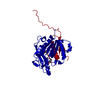


 PDBj
PDBj

 Assembly


 Mass: 96.063 Da / Num. of mol.: 1 / Source method: obtained synthetically / Formula: SO4
 Mass: 18.015 Da / Num. of mol.: 78 / Source method: isolated from a natural source / Formula: H2O
 Sample preparation
 / Beamline: BL1-5 / Wavelength: 0.918381, 0.979094,0.978532
 Processing