 Movie
Movie Controller

[English] 日本語
 Yorodumi
- PDB-5zol: Crystal structure of a three sites mutantion of FSAA complexed wi... -
+ Open data

- Basic information
| Entry | Database: PDB / ID: 5zol | ||||||
|---|---|---|---|---|---|---|---|
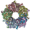
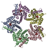
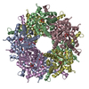
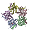
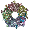
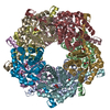
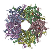
| Title | Crystal structure of a three sites mutantion of FSAA complexed with HA and product | ||||||
 Components Components | Fructose-6-phosphate aldolase 1 | ||||||
 Keywords Keywords | LYASE / Fructose-6-phosphate aldolase / donor / acceptor / mutant / flexible | ||||||
| Function / homology |  Function and homology information Function and homology informationketone catabolic process / fructose 6-phosphate aldolase activity / Lyases; Carbon-carbon lyases; Aldehyde-lyases / fructose metabolic process / identical protein binding / cytoplasm Similarity search - Function | ||||||
| Biological species |  | ||||||
| Method |  X-RAY DIFFRACTION / SYNCHROTRON / Resolution: 2.172 Å X-RAY DIFFRACTION / SYNCHROTRON / Resolution: 2.172 Å | ||||||
 Authors Authors | Wu, L. / Yang, X.H. / Yu, H.W. / Zhou, J.H. | ||||||
 Citation Citation | Journal: Chem.Commun.(Camb.) / Year: 2020 Title: The engineering of decameric d-fructose-6-phosphate aldolase A by combinatorial modulation of inter- and intra-subunit interactions. Authors: Yang, X. / Wu, L. / Li, A. / Ye, L. / Zhou, J. / Yu, H. | ||||||
| History |
|
- Structure visualization
| Structure viewer | Molecule: MolmilJmol/JSmol |
|---|
- Downloads & links
-Download
| PDBx/mmCIF format | 5zol.cif.gz | 437.6 KB | Display | PDBx/mmCIF format |
|---|---|---|---|---|
| PDB format | pdb5zol.ent.gz | 356 KB | Display | PDB format |
| PDBx/mmJSON format | 5zol.json.gz | Tree view | PDBx/mmJSON format | |
| Others |  Other downloads Other downloads |
-Validation report
| Arichive directory | https://data.pdbj.org/pub/pdb/validation_reports/zo/5zolftp://data.pdbj.org/pub/pdb/validation_reports/zo/5zol | HTTPS FTP |
|---|
-Related structure data
| Similar structure data |
|---|


-Links
 PDBj
PDBj- Assembly
| Deposited unit | 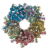
| |||||||||
|---|---|---|---|---|---|---|---|---|---|---|
| 1 |
| |||||||||
| Unit cell |
| |||||||||
| Components on special symmetry positions |
|
-Components
-Protein , 1 types, 10 molecules ABCDEFGHIJ
| #1: Protein | Mass: 26928.037 Da / Num. of mol.: 10 / Mutation: T31I/T59Q/Q195I Source method: isolated from a genetically manipulated source Source: (gene. exp.) Strain: K12 / Gene: fsaA, fsa, mipB, ybiZ, b0825, JW5109 / Production host: References: UniProt: P78055, Lyases; Carbon-carbon lyases; Aldehyde-lyases |
|---|
-Non-polymers , 6 types, 946 molecules 


| #2: Chemical | ChemComp-4Y8 /  Mass: 74.079 Da / Num. of mol.: 10 Mass: 74.079 Da / Num. of mol.: 10Source method: isolated from a genetically manipulated source Formula: C3H6O2 #3: Chemical | ChemComp-LW2 / (  Mass: 186.228 Da / Num. of mol.: 9 Mass: 186.228 Da / Num. of mol.: 9Source method: isolated from a genetically manipulated source Formula: C8H10O3S #4: Chemical | ChemComp-CL /  Mass: 35.453 Da / Num. of mol.: 4 / Source method: obtained synthetically / Formula: Cl Mass: 35.453 Da / Num. of mol.: 4 / Source method: obtained synthetically / Formula: Cl#5: Chemical | ChemComp-SO4 /  Mass: 96.063 Da / Num. of mol.: 9 / Source method: obtained synthetically / Formula: SO4 Mass: 96.063 Da / Num. of mol.: 9 / Source method: obtained synthetically / Formula: SO4#6: Chemical | ChemComp-LW1 / |  Mass: 112.150 Da / Num. of mol.: 1 / Source method: obtained synthetically / Formula: C5H4OS Mass: 112.150 Da / Num. of mol.: 1 / Source method: obtained synthetically / Formula: C5H4OS#7: Water | ChemComp-HOH / | Mass: 18.015 Da / Num. of mol.: 913 / Source method: isolated from a natural source / Formula: H2O |
|---|
-Details
| Has protein modification | Y |
|---|
-Experimental details
-Experiment
| Experiment | Method: X-RAY DIFFRACTION / Number of used crystals: 1 |
|---|
- Sample preparation
| Crystal | Density Matthews: 2.81 Å3/Da / Density % sol: 56.19 % |
|---|---|
| Crystal grow | Temperature: 293 K / Method: vapor diffusion, sitting drop Details: 0.1M Hepes pH7.2, 18% PEG 4000, 10% isopropyl alcohol, 0.2M ammonium sulfate, 0.01M disodium malonate |
-Data collection
| Diffraction | Mean temperature: 100 K |
|---|---|
| Diffraction source | Source: SYNCHROTRON / Site: SSRF  / Beamline: BL19U1 / Wavelength: 0.97853 Å / Beamline: BL19U1 / Wavelength: 0.97853 Å |
| Detector | Type: DECTRIS PILATUS3 S 6M / Detector: PIXEL / Date: Jan 5, 2018 |
| Radiation | Protocol: SINGLE WAVELENGTH / Monochromatic (M) / Laue (L): M / Scattering type: x-ray |
| Radiation wavelength | Wavelength: 0.97853 Å / Relative weight: 1 |
| Reflection | Resolution: 2.17→50 Å / Num. obs: 161347 / % possible obs: 99.7 % / Redundancy: 16.3 % / Net I/σ(I): 2 |
| Reflection shell | Resolution: 2.17→2.21 Å / Redundancy: 15.8 % / % possible all: 100 |
- Processing
| Software |
| |||||||||||||||||||||||||||||||||||||||||||||||||||||||||||||||||||||||||||||||||||||||||||||||||||||||||||||||||||||||||||||||||||||||||||||||||||||||||||||||||||||||||||||||||||||||||||||||||||||||||||||||||||||||||
|---|---|---|---|---|---|---|---|---|---|---|---|---|---|---|---|---|---|---|---|---|---|---|---|---|---|---|---|---|---|---|---|---|---|---|---|---|---|---|---|---|---|---|---|---|---|---|---|---|---|---|---|---|---|---|---|---|---|---|---|---|---|---|---|---|---|---|---|---|---|---|---|---|---|---|---|---|---|---|---|---|---|---|---|---|---|---|---|---|---|---|---|---|---|---|---|---|---|---|---|---|---|---|---|---|---|---|---|---|---|---|---|---|---|---|---|---|---|---|---|---|---|---|---|---|---|---|---|---|---|---|---|---|---|---|---|---|---|---|---|---|---|---|---|---|---|---|---|---|---|---|---|---|---|---|---|---|---|---|---|---|---|---|---|---|---|---|---|---|---|---|---|---|---|---|---|---|---|---|---|---|---|---|---|---|---|---|---|---|---|---|---|---|---|---|---|---|---|---|---|---|---|---|---|---|---|---|---|---|---|---|---|---|---|---|---|---|---|---|
| Refinement | Resolution: 2.172→22.7 Å / SU ML: 0.28 / Cross valid method: FREE R-VALUE / σ(F): 1.35 / Phase error: 24.43
| |||||||||||||||||||||||||||||||||||||||||||||||||||||||||||||||||||||||||||||||||||||||||||||||||||||||||||||||||||||||||||||||||||||||||||||||||||||||||||||||||||||||||||||||||||||||||||||||||||||||||||||||||||||||||
| Solvent computation | Shrinkage radii: 0.9 Å / VDW probe radii: 1.11 Å | |||||||||||||||||||||||||||||||||||||||||||||||||||||||||||||||||||||||||||||||||||||||||||||||||||||||||||||||||||||||||||||||||||||||||||||||||||||||||||||||||||||||||||||||||||||||||||||||||||||||||||||||||||||||||
| Refinement step | Cycle: LAST / Resolution: 2.172→22.7 Å
| |||||||||||||||||||||||||||||||||||||||||||||||||||||||||||||||||||||||||||||||||||||||||||||||||||||||||||||||||||||||||||||||||||||||||||||||||||||||||||||||||||||||||||||||||||||||||||||||||||||||||||||||||||||||||
| Refine LS restraints |
| |||||||||||||||||||||||||||||||||||||||||||||||||||||||||||||||||||||||||||||||||||||||||||||||||||||||||||||||||||||||||||||||||||||||||||||||||||||||||||||||||||||||||||||||||||||||||||||||||||||||||||||||||||||||||
| LS refinement shell |
|
