 Movie
Movie Controller


+ Open data

- Basic information
| Entry | Database: PDB / ID: 5l0f | ||||||
|---|---|---|---|---|---|---|---|
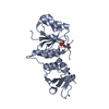
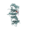
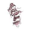
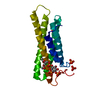
| Title | Human metavinculin quadruple mutant (residues 959-1134) | ||||||
 Components Components | Vinculin | ||||||
 Keywords Keywords | CELL ADHESION / 5-Helix bundle / cytoskelatal protein / phospholipids / structural protein | ||||||
| Function / homology |  Function and homology information Function and homology informationregulation of protein localization to adherens junction / outer dense plaque of desmosome / inner dense plaque of desmosome / podosome ring / terminal web / cell-substrate junction / epithelial cell-cell adhesion / zonula adherens / fascia adherens / dystroglycan binding ...regulation of protein localization to adherens junction / outer dense plaque of desmosome / inner dense plaque of desmosome / podosome ring / terminal web / cell-substrate junction / epithelial cell-cell adhesion / zonula adherens / fascia adherens / dystroglycan binding / alpha-catenin binding / cell-cell contact zone / apical junction assembly / costamere / regulation of establishment of endothelial barrier / axon extension / protein localization to cell surface / adherens junction assembly / lamellipodium assembly / regulation of focal adhesion assembly / maintenance of blood-brain barrier / brush border / Smooth Muscle Contraction / negative regulation of cell migration / Turbulent (oscillatory, disturbed) flow shear stress activates signaling by PIEZO1 and integrins in endothelial cells / morphogenesis of an epithelium / cell projection / cell-matrix adhesion / adherens junction / Signaling by high-kinase activity BRAF mutants / MAP2K and MAPK activation / beta-catenin binding / sarcolemma / platelet aggregation / specific granule lumen / Signaling by RAF1 mutants / Signaling by moderate kinase activity BRAF mutants / Paradoxical activation of RAF signaling by kinase inactive BRAF / Signaling downstream of RAS mutants / cell-cell junction / Signaling by BRAF and RAF1 fusions / Signaling by ALK fusions and activated point mutants / Platelet degranulation / extracellular vesicle / actin binding / secretory granule lumen / High laminar flow shear stress activates signaling by PIEZO1 and PECAM1:CDH5:KDR in endothelial cells / ficolin-1-rich granule lumen / molecular adaptor activity / cytoskeleton / cell adhesion / cadherin binding / membrane raft / focal adhesion / ubiquitin protein ligase binding / Neutrophil degranulation / structural molecule activity / protein-containing complex / extracellular exosome / extracellular region / plasma membrane / cytoplasm / cytosol Similarity search - Function | ||||||
| Biological species |  Homo sapiens (human) Homo sapiens (human) | ||||||
| Method |  X-RAY DIFFRACTION / SYNCHROTRON / MOLECULAR REPLACEMENT / Resolution: 2.76 Å X-RAY DIFFRACTION / SYNCHROTRON / MOLECULAR REPLACEMENT / Resolution: 2.76 Å | ||||||
 Authors Authors | Chinthalapudi, K. / Izard, T. | ||||||
 Citation Citation | Journal: Proc.Natl.Acad.Sci.USA / Year: 2016 Title: Differential lipid binding of vinculin isoforms promotes quasi-equivalent dimerization. Authors: Chinthalapudi, K. / Rangarajan, E.S. / Brown, D.T. / Izard, T. | ||||||
| History |
|
- Structure visualization
| Structure viewer | Molecule: MolmilJmol/JSmol |
|---|
- Downloads & links
-Download
| PDBx/mmCIF format | 5l0f.cif.gz | 86.9 KB | Display | PDBx/mmCIF format |
|---|---|---|---|---|
| PDB format | pdb5l0f.ent.gz | 65.6 KB | Display | PDB format |
| PDBx/mmJSON format | 5l0f.json.gz | Tree view | PDBx/mmJSON format | |
| Others |  Other downloads Other downloads |
-Validation report
| Arichive directory | https://data.pdbj.org/pub/pdb/validation_reports/l0/5l0fftp://data.pdbj.org/pub/pdb/validation_reports/l0/5l0f | HTTPS FTP |
|---|
-Related structure data
| Related structure data |  5l0cC  5l0dC  5l0gC  5l0hC  5l0iC  5l0jC 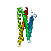 3myiS S: Starting model for refinement C: citing same article ( |
|---|---|
| Similar structure data |


-Links
 PDBj
PDBj


- Assembly
| Deposited unit | 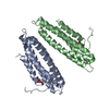
| ||||||||
|---|---|---|---|---|---|---|---|---|---|
| 1 | 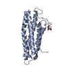
| ||||||||
| 2 | 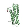
| ||||||||
| Unit cell |
|
-Components
| #1: Protein | Mass: 19531.453 Da / Num. of mol.: 2 / Fragment: UNP Residues 959-1134 / Mutation: R975Q, K979Q, R1107Q, R1128Q Source method: isolated from a genetically manipulated source Source: (gene. exp.) Homo sapiens (human) / Gene: VCL / Plasmid: PET28 / Production host:  #2: Chemical |   Mass: 92.094 Da / Num. of mol.: 2 / Source method: obtained synthetically / Formula: C3H8O3 Mass: 92.094 Da / Num. of mol.: 2 / Source method: obtained synthetically / Formula: C3H8O3#3: Water | ChemComp-HOH / |  Mass: 18.015 Da / Num. of mol.: 287 / Source method: isolated from a natural source / Formula: H2O Mass: 18.015 Da / Num. of mol.: 287 / Source method: isolated from a natural source / Formula: H2O |
|---|
-Experimental details
-Experiment
| Experiment | Method: X-RAY DIFFRACTION / Number of used crystals: 1 |
|---|
- Sample preparation
| Crystal | Density Matthews: 2.52 Å3/Da / Density % sol: 51.2 % |
|---|---|
| Crystal grow | Temperature: 294 K / Method: vapor diffusion, hanging drop / pH: 8.8 Details: 20 mM citric acid, 80 mM Bis-Tris propane (pH 8.8), and 14 to 16% (w/v) PEG 3350 |
-Data collection
| Diffraction | Mean temperature: 100 K |
|---|---|
| Diffraction source | Source: SYNCHROTRON / Site: APS  / Beamline: 22-BM / Wavelength: 1 Å / Beamline: 22-BM / Wavelength: 1 Å |
| Detector | Type: MARMOSAIC 225 mm CCD / Detector: CCD / Date: Jul 23, 2014 |
| Radiation | Monochromator: double crystal Si(111) / Protocol: SINGLE WAVELENGTH / Monochromatic (M) / Laue (L): M / Scattering type: x-ray |
| Radiation wavelength | Wavelength: 1 Å / Relative weight: 1 |
| Reflection | Resolution: 2.76→60.79 Å / Num. obs: 10731 / % possible obs: 99.8 % / Redundancy: 9.6 % / Biso Wilson estimate: 40.61 Å2 / CC1/2: 0.999 / Rpim(I) all: 0.022 / Net I/σ(I): 28.1 |
| Reflection shell | Resolution: 2.76→2.91 Å / Redundancy: 9.1 % / Mean I/σ(I) obs: 13 / CC1/2: 0.995 / % possible all: 99.7 |
- Processing
| Software |
| ||||||||||||||||||||||||||||||||||||||||||||||||||||||||||||||||||||||||||||||||||||||||||||||||||||||||||||||||||
|---|---|---|---|---|---|---|---|---|---|---|---|---|---|---|---|---|---|---|---|---|---|---|---|---|---|---|---|---|---|---|---|---|---|---|---|---|---|---|---|---|---|---|---|---|---|---|---|---|---|---|---|---|---|---|---|---|---|---|---|---|---|---|---|---|---|---|---|---|---|---|---|---|---|---|---|---|---|---|---|---|---|---|---|---|---|---|---|---|---|---|---|---|---|---|---|---|---|---|---|---|---|---|---|---|---|---|---|---|---|---|---|---|---|---|---|
| Refinement | Method to determine structure: MOLECULAR REPLACEMENT Starting model: 3MYI Resolution: 2.76→60.79 Å / Cor.coef. Fo:Fc: 0.9444 / Cor.coef. Fo:Fc free: 0.9199 / Cross valid method: THROUGHOUT / σ(F): 0 / SU Rfree Blow DPI: 0.381
| ||||||||||||||||||||||||||||||||||||||||||||||||||||||||||||||||||||||||||||||||||||||||||||||||||||||||||||||||||
| Displacement parameters | Biso mean: 26.93 Å2
| ||||||||||||||||||||||||||||||||||||||||||||||||||||||||||||||||||||||||||||||||||||||||||||||||||||||||||||||||||
| Refine analyze | Luzzati coordinate error obs: 0.265 Å | ||||||||||||||||||||||||||||||||||||||||||||||||||||||||||||||||||||||||||||||||||||||||||||||||||||||||||||||||||
| Refinement step | Cycle: 1 / Resolution: 2.76→60.79 Å
| ||||||||||||||||||||||||||||||||||||||||||||||||||||||||||||||||||||||||||||||||||||||||||||||||||||||||||||||||||
| Refine LS restraints |
| ||||||||||||||||||||||||||||||||||||||||||||||||||||||||||||||||||||||||||||||||||||||||||||||||||||||||||||||||||
| LS refinement shell | Resolution: 2.76→3.08 Å / Total num. of bins used: 5
|
