Protocol: SINGLE WAVELENGTH / Monochromatic (M) / Laue (L): M / Scattering type: x-ray
Radiation wavelength
Wavelength: 1.008 Å / Relative weight: 1
Reflection
Resolution: 1.05→91.87 Å / Num. obs: 144393 / % possible obs: 88.8 % / Redundancy: 5 % / Biso Wilson estimate: 5.7 Å2 / Rmerge(I) obs: 0.079 / Rpim(I) all: 0.034 / Net I/σ(I): 11.9
Reflection shell
Diffraction-ID: 1
Resolution (Å)
Redundancy (%)
Rmerge(I) obs
Mean I/σ(I) obs
Num. unique obs
Rpim(I) all
% possible all
1.05-1.11
2
0.336
2.1
10354
0.308
43.8
3.32-37.43
7.2
0.054
31.1
5234
0.022
99.9
-
Processing
Software
Name
Version
Classification
REFMAC
5.8.0124
refinement
XDS
datareduction
SCALA
datascaling
SHELXD
phasing
Refinement
Method to determine structure: MOLECULAR REPLACEMENT Starting model: Se-SAD model Resolution: 1.05→91.87 Å / Cor.coef. Fo:Fc: 0.982 / Cor.coef. Fo:Fc free: 0.975 / SU B: 0.738 / SU ML: 0.016 / Cross valid method: THROUGHOUT / ESU R: 0.025 / ESU R Free: 0.026 / Stereochemistry target values: MAXIMUM LIKELIHOOD / Details: HYDROGENS HAVE BEEN ADDED IN THE RIDING POSITIONS
Rfactor
Num. reflection
% reflection
Selection details
Rfree
0.14252
7242
5 %
RANDOM
Rwork
0.11711
-
-
-
obs
0.11839
137073
88.7 %
-
Solvent computation
Ion probe radii: 0.8 Å / Shrinkage radii: 0.8 Å / VDW probe radii: 1.2 Å / Solvent model: MASK
 Movie
Movie Controller

 Yorodumi
 Open data
 Basic information
 Components
 Keywords
 Function and homology information
 X-RAY DIFFRACTION /
  Authors
 Belgium, 1items
  Citation
 Structure visualization
 Downloads & links
 Other downloads


 PDBj
PDBj Assembly


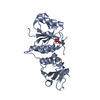
 Mass: 96.063 Da / Num. of mol.: 1 / Source method: obtained synthetically / Formula: SO4
 Mass: 18.015 Da / Num. of mol.: 646 / Source method: isolated from a natural source / Formula: H2O
 Sample preparation
 / Beamline: ID29 / Wavelength: 1.008 Å
 Processing