- PDB-3ltl: Crystal structure of human BIG1 Sec7 domain -
+
Open data
ID or keywords:
Loading...
-
Basic information
Entry
Database: PDB / ID: 3ltl
Title
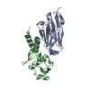
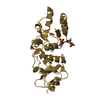
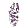
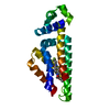
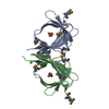
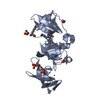
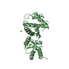
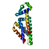
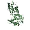
Crystal structure of human BIG1 Sec7 domain
Components
Brefeldin A-inhibited guanine nucleotide-exchange protein 1
Keywords
SIGNALING PROTEIN / all alpha / Guanine-nucleotide releasing factor
Function / homology
Function and homology information
endomembrane system organization / negative regulation of actin filament polymerization / negative regulation of GTPase activity / glycoprotein biosynthetic process / small nuclear ribonucleoprotein complex / regulation of ARF protein signal transduction / positive regulation of wound healing / myosin binding / regulation of establishment of cell polarity / protein kinase A regulatory subunit binding ...endomembrane system organization / negative regulation of actin filament polymerization / negative regulation of GTPase activity / glycoprotein biosynthetic process / small nuclear ribonucleoprotein complex / regulation of ARF protein signal transduction / positive regulation of wound healing / myosin binding / regulation of establishment of cell polarity / protein kinase A regulatory subunit binding / Golgi organization / exocytosis / guanyl-nucleotide exchange factor activity / trans-Golgi network / nuclear matrix / neuron projection development / protein transport / positive regulation of phosphatidylinositol 3-kinase/protein kinase B signal transduction / Golgi membrane / nucleolus / perinuclear region of cytoplasm / Golgi apparatus / nucleoplasm / cytosol Similarity search - Function
In the structure databanks used in Yorodumi, some data are registered as the other names, "COVID-19 virus" and "2019-nCoV". Here are the details of the virus and the list of structure data.
Jan 31, 2019. EMDB accession codes are about to change! (news from PDBe EMDB page)
EMDB accession codes are about to change! (news from PDBe EMDB page)
The allocation of 4 digits for EMDB accession codes will soon come to an end. Whilst these codes will remain in use, new EMDB accession codes will include an additional digit and will expand incrementally as the available range of codes is exhausted. The current 4-digit format prefixed with “EMD-” (i.e. EMD-XXXX) will advance to a 5-digit format (i.e. EMD-XXXXX), and so on. It is currently estimated that the 4-digit codes will be depleted around Spring 2019, at which point the 5-digit format will come into force.
The EM Navigator/Yorodumi systems omit the EMD- prefix.
Related info.:Q: What is EMD? / ID/Accession-code notation in Yorodumi/EM Navigator
Yorodumi is a browser for structure data from EMDB, PDB, SASBDB, etc.
This page is also the successor to EM Navigator detail page, and also detail information page/front-end page for Omokage search.
The word "yorodu" (or yorozu) is an old Japanese word meaning "ten thousand". "mi" (miru) is to see.
Related info.:EMDB / PDB / SASBDB / Comparison of 3 databanks / Yorodumi Search / Aug 31, 2016. New EM Navigator & Yorodumi / Yorodumi Papers / Jmol/JSmol / Function and homology information / Changes in new EM Navigator and Yorodumi
 Movie
Movie Controller


 Open data
 Basic information
 Components
 Keywords
 Function and homology information
 Homo sapiens (human)
 X-RAY DIFFRACTION /
  Authors
 Citation
 Structure visualization
 Downloads & links
 Other downloads


 PDBj
PDBj

 Assembly
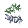


 Mass: 40.078 Da / Num. of mol.: 2 / Source method: obtained synthetically / Formula: Ca
 Mass: 60.052 Da / Num. of mol.: 1 / Source method: obtained synthetically / Formula: C2H4O2
 Mass: 18.015 Da / Num. of mol.: 73 / Source method: isolated from a natural source / Formula: H2O
 Sample preparation
 / Beamline: ID14-1 / Wavelength: 0.934 Å
 Processing