 Movie
Movie Controller


+ Open data

- Basic information
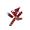
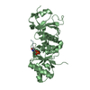
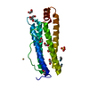
| Entry | Database: PDB / ID: 5v89 | ||||||
|---|---|---|---|---|---|---|---|
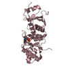
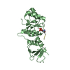
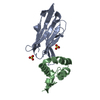
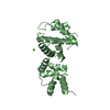
| Title | Structure of DCN4 PONY domain bound to CUL1 WHB | ||||||
 Components Components |
| ||||||
 Keywords Keywords | Ligase / protein binding / E3 Ligase / Ligase - protein binding complex | ||||||
| Function / homology |  Function and homology information Function and homology informationpositive regulation of protein neddylation / ubiquitin-like protein binding / Parkin-FBXW7-Cul1 ubiquitin ligase complex / cullin-RING ubiquitin ligase complex / Loss of Function of FBXW7 in Cancer and NOTCH1 Signaling / protein neddylation / ubiquitin conjugating enzyme binding / SCF ubiquitin ligase complex / SCF-dependent proteasomal ubiquitin-dependent protein catabolic process / Prolactin receptor signaling ...positive regulation of protein neddylation / ubiquitin-like protein binding / Parkin-FBXW7-Cul1 ubiquitin ligase complex / cullin-RING ubiquitin ligase complex / Loss of Function of FBXW7 in Cancer and NOTCH1 Signaling / protein neddylation / ubiquitin conjugating enzyme binding / SCF ubiquitin ligase complex / SCF-dependent proteasomal ubiquitin-dependent protein catabolic process / Prolactin receptor signaling / ubiquitin ligase complex scaffold activity / cullin family protein binding / protein monoubiquitination / ubiquitin ligase complex / Nuclear events stimulated by ALK signaling in cancer / protein K48-linked ubiquitination / intrinsic apoptotic signaling pathway / animal organ morphogenesis / Regulation of BACH1 activity / MAP3K8 (TPL2)-dependent MAPK1/3 activation / G1/S transition of mitotic cell cycle / SCF-beta-TrCP mediated degradation of Emi1 / NIK-->noncanonical NF-kB signaling / Dectin-1 mediated noncanonical NF-kB signaling / Degradation of CRY and PER proteins / Activation of NF-kappaB in B cells / Iron uptake and transport / Degradation of GLI1 by the proteasome / GSK3B and BTRC:CUL1-mediated-degradation of NFE2L2 / Negative regulation of NOTCH4 signaling / FBXL7 down-regulates AURKA during mitotic entry and in early mitosis / Degradation of GLI2 by the proteasome / GLI3 is processed to GLI3R by the proteasome / Ubiquitin-Mediated Degradation of Phosphorylated Cdc25A / NOTCH1 Intracellular Domain Regulates Transcription / Degradation of beta-catenin by the destruction complex / Constitutive Signaling by NOTCH1 PEST Domain Mutants / Constitutive Signaling by NOTCH1 HD+PEST Domain Mutants / CLEC7A (Dectin-1) signaling / SCF(Skp2)-mediated degradation of p27/p21 / FCERI mediated NF-kB activation / Interleukin-1 signaling / Orc1 removal from chromatin / Cyclin D associated events in G1 / Regulation of RUNX2 expression and activity / Regulation of PLK1 Activity at G2/M Transition / Downstream TCR signaling / Antigen processing: Ubiquitination & Proteasome degradation / Neddylation / cellular response to oxidative stress / proteasome-mediated ubiquitin-dependent protein catabolic process / protein-macromolecule adaptor activity / positive regulation of canonical NF-kappaB signal transduction / cell population proliferation / protein ubiquitination / ubiquitin protein ligase binding / nucleoplasm / nucleus / plasma membrane / cytosol / cytoplasm Similarity search - Function | ||||||
| Biological species |  Homo sapiens (human) Homo sapiens (human) | ||||||
| Method |  X-RAY DIFFRACTION / SYNCHROTRON / MOLECULAR REPLACEMENT / Resolution: 1.55 Å X-RAY DIFFRACTION / SYNCHROTRON / MOLECULAR REPLACEMENT / Resolution: 1.55 Å | ||||||
 Authors Authors | Guy, R.K. / Schulman, B.A. / Scott, D.C. / Hammill, J.T. | ||||||
| Funding support |  United States, 1items United States, 1items
| ||||||
 Citation Citation | Journal: Nat. Chem. Biol. / Year: 2017 Title: Blocking an N-terminal acetylation-dependent protein interaction inhibits an E3 ligase. Authors: Scott, D.C. / Hammill, J.T. / Min, J. / Rhee, D.Y. / Connelly, M. / Sviderskiy, V.O. / Bhasin, D. / Chen, Y. / Ong, S.S. / Chai, S.C. / Goktug, A.N. / Huang, G. / Monda, J.K. / Low, J. / ...Authors: Scott, D.C. / Hammill, J.T. / Min, J. / Rhee, D.Y. / Connelly, M. / Sviderskiy, V.O. / Bhasin, D. / Chen, Y. / Ong, S.S. / Chai, S.C. / Goktug, A.N. / Huang, G. / Monda, J.K. / Low, J. / Kim, H.S. / Paulo, J.A. / Cannon, J.R. / Shelat, A.A. / Chen, T. / Kelsall, I.R. / Alpi, A.F. / Pagala, V. / Wang, X. / Peng, J. / Singh, B. / Harper, J.W. / Schulman, B.A. / Guy, R.K. | ||||||
| History |
|
- Structure visualization
| Structure viewer | Molecule: MolmilJmol/JSmol |
|---|
- Downloads & links
-Download
| PDBx/mmCIF format | 5v89.cif.gz | 172 KB | Display | PDBx/mmCIF format |
|---|---|---|---|---|
| PDB format | pdb5v89.ent.gz | 136 KB | Display | PDB format |
| PDBx/mmJSON format | 5v89.json.gz | Tree view | PDBx/mmJSON format | |
| Others |  Other downloads Other downloads |
-Validation report
| Arichive directory | https://data.pdbj.org/pub/pdb/validation_reports/v8/5v89ftp://data.pdbj.org/pub/pdb/validation_reports/v8/5v89 | HTTPS FTP |
|---|
-Related structure data
| Related structure data | 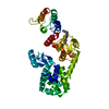 5v83C  5v86C  5v88C  3tduS C: citing same article ( S: Starting model for refinement |
|---|---|
| Similar structure data |
-Links
 PDBj
PDBj


- Assembly
| Deposited unit | 
| ||||||||
|---|---|---|---|---|---|---|---|---|---|
| 1 | 
| ||||||||
| 2 | 
| ||||||||
| Unit cell |
| ||||||||
| Details | Monomer as determined by gel filtration |
-Components
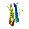
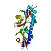
| #1: Protein | Mass: 22756.834 Da / Num. of mol.: 1 / Fragment: residues 102-292 Source method: isolated from a genetically manipulated source Source: (gene. exp.) Homo sapiens (human) / Gene: DCUN1D4, KIAA0276 / Plasmid: pGEX / Production host:  |
|---|---|
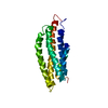
| #2: Protein | Mass: 9015.699 Da / Num. of mol.: 1 / Fragment: WHB subdomain residues 702-776 Source method: isolated from a genetically manipulated source Source: (gene. exp.) Homo sapiens (human) / Gene: CUL1 / Plasmid: pGEX / Production host: |
| #3: Water | ChemComp-HOH /  Mass: 18.015 Da / Num. of mol.: 312 / Source method: isolated from a natural source / Formula: H2O Mass: 18.015 Da / Num. of mol.: 312 / Source method: isolated from a natural source / Formula: H2O |
-Experimental details
-Experiment
| Experiment | Method: X-RAY DIFFRACTION / Number of used crystals: 1 |
|---|
- Sample preparation
| Crystal | Density Matthews: 2.17 Å3/Da / Density % sol: 43.42 % Description: The authors state that the density surrounding the CUL1WHB subdomain in this structure was relatively weak, presumably due to its lack of participation in forming crystal contacts. ...Description: The authors state that the density surrounding the CUL1WHB subdomain in this structure was relatively weak, presumably due to its lack of participation in forming crystal contacts. Therefore we modeled and built this chain largely from a previous high resolution and high quality structure of CUL1WHB, 3TDU. |
|---|---|
| Crystal grow | Temperature: 277 K / Method: vapor diffusion, hanging drop / pH: 6.5 Details: 25% PEG3350, 0.2M LiSO4, 10mM TCEP, 10mM Bis-Tris Propane |
-Data collection
| Diffraction | Mean temperature: 100 K | |||||||||||||||||||||||||||||||||||||||||||||||||||||||||||||||||||||||||||||
|---|---|---|---|---|---|---|---|---|---|---|---|---|---|---|---|---|---|---|---|---|---|---|---|---|---|---|---|---|---|---|---|---|---|---|---|---|---|---|---|---|---|---|---|---|---|---|---|---|---|---|---|---|---|---|---|---|---|---|---|---|---|---|---|---|---|---|---|---|---|---|---|---|---|---|---|---|---|---|
| Diffraction source | Source: SYNCHROTRON / Site: APS / Beamline: 22-ID / Wavelength: 1 Å | |||||||||||||||||||||||||||||||||||||||||||||||||||||||||||||||||||||||||||||
| Detector | Type: MARMOSAIC 300 mm CCD / Detector: CCD / Date: Jun 15, 2012 | |||||||||||||||||||||||||||||||||||||||||||||||||||||||||||||||||||||||||||||
| Radiation | Protocol: SINGLE WAVELENGTH / Monochromatic (M) / Laue (L): M / Scattering type: x-ray | |||||||||||||||||||||||||||||||||||||||||||||||||||||||||||||||||||||||||||||
| Radiation wavelength | Wavelength: 1 Å / Relative weight: 1 | |||||||||||||||||||||||||||||||||||||||||||||||||||||||||||||||||||||||||||||
| Reflection | Resolution: 1.55→50 Å / Num. obs: 36927 / % possible obs: 93.4 % / Redundancy: 3.5 % / Rmerge(I) obs: 0.072 / Χ2: 1.061 / Net I/σ(I): 7.7 | |||||||||||||||||||||||||||||||||||||||||||||||||||||||||||||||||||||||||||||
| Reflection shell |
|
- Processing
| Software |
| ||||||||||||||||||||||||||||||||||||||||||||||||||||||||||||||||||||||||||||||||||||||||||||||||||
|---|---|---|---|---|---|---|---|---|---|---|---|---|---|---|---|---|---|---|---|---|---|---|---|---|---|---|---|---|---|---|---|---|---|---|---|---|---|---|---|---|---|---|---|---|---|---|---|---|---|---|---|---|---|---|---|---|---|---|---|---|---|---|---|---|---|---|---|---|---|---|---|---|---|---|---|---|---|---|---|---|---|---|---|---|---|---|---|---|---|---|---|---|---|---|---|---|---|---|---|
| Refinement | Method to determine structure: MOLECULAR REPLACEMENT Starting model: 3TDU.pdb Resolution: 1.55→23.917 Å / SU ML: 0.16 / Cross valid method: FREE R-VALUE / σ(F): 1.34 / Phase error: 19.42
| ||||||||||||||||||||||||||||||||||||||||||||||||||||||||||||||||||||||||||||||||||||||||||||||||||
| Solvent computation | Shrinkage radii: 0.9 Å / VDW probe radii: 1.11 Å | ||||||||||||||||||||||||||||||||||||||||||||||||||||||||||||||||||||||||||||||||||||||||||||||||||
| Displacement parameters | Biso max: 109.85 Å2 / Biso mean: 28.4826 Å2 / Biso min: 7.28 Å2 | ||||||||||||||||||||||||||||||||||||||||||||||||||||||||||||||||||||||||||||||||||||||||||||||||||
| Refinement step | Cycle: final / Resolution: 1.55→23.917 Å
| ||||||||||||||||||||||||||||||||||||||||||||||||||||||||||||||||||||||||||||||||||||||||||||||||||
| Refine LS restraints |
| ||||||||||||||||||||||||||||||||||||||||||||||||||||||||||||||||||||||||||||||||||||||||||||||||||
| LS refinement shell | Refine-ID: X-RAY DIFFRACTION / Rfactor Rfree error: 0 / Total num. of bins used: 13
|
