 Movie
Movie Controller

[English] 日本語
 Yorodumi
- PDB-5kcn: Crystal Structure of full-length LpoA from Haemophilus influenzae... -
+ Open data

- Basic information
| Entry | Database: PDB / ID: 5kcn | |||||||||
|---|---|---|---|---|---|---|---|---|---|---|
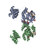
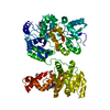
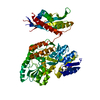
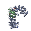
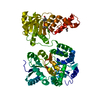
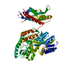
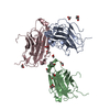
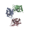
| Title | Crystal Structure of full-length LpoA from Haemophilus influenzae at 1.97 angstrom resolution | |||||||||
 Components Components | Penicillin-binding protein activator LpoA | |||||||||
 Keywords Keywords | BIOSYNTHETIC PROTEIN / Outer membrane lipoprotein / PBP1A / transpeptidase. peptidoglycan | |||||||||
| Function / homology |  Function and homology information Function and homology informationperiplasmic side of cell outer membrane / peptidoglycan biosynthetic process / enzyme regulator activity / regulation of cell shape Similarity search - Function | |||||||||
| Biological species |  Haemophilus influenzae (bacteria) Haemophilus influenzae (bacteria) | |||||||||
| Method |  X-RAY DIFFRACTION / SYNCHROTRON / MOLECULAR REPLACEMENT / Resolution: 1.965 Å X-RAY DIFFRACTION / SYNCHROTRON / MOLECULAR REPLACEMENT / Resolution: 1.965 Å | |||||||||
 Authors Authors | Sathiyamoorthy, K. / Saper, M.A. | |||||||||
| Funding support |  United States, 2items United States, 2items
| |||||||||
 Citation Citation | Journal: J. Biol. Chem. / Year: 2017 Title: Structural analyses of the Haemophilus influenzae peptidoglycan synthase activator LpoA suggest multiple conformations in solution. Authors: Sathiyamoorthy, K. / Vijayalakshmi, J. / Tirupati, B. / Fan, L. / Saper, M.A. #1: Journal: Proteins / Year: 2008Title: Structure of YraM, a protein essential for growth of Haemophilus influenzae. Authors: Vijayalakshmi, J. / Akerley, B.J. / Saper, M.A. | |||||||||
| History |
|
- Structure visualization
| Structure viewer | Molecule: MolmilJmol/JSmol |
|---|
- Downloads & links
-Download
| PDBx/mmCIF format | 5kcn.cif.gz | 322.3 KB | Display | PDBx/mmCIF format |
|---|---|---|---|---|
| PDB format | pdb5kcn.ent.gz | 264.8 KB | Display | PDB format |
| PDBx/mmJSON format | 5kcn.json.gz | Tree view | PDBx/mmJSON format | |
| Others |  Other downloads Other downloads |
-Validation report
| Arichive directory | https://data.pdbj.org/pub/pdb/validation_reports/kc/5kcnftp://data.pdbj.org/pub/pdb/validation_reports/kc/5kcn | HTTPS FTP |
|---|
-Related structure data
| Related structure data |  4p29SC  5vatC  5vbgC  3ckmS S: Starting model for refinement C: citing same article ( |
|---|---|
| Similar structure data |


-Links
 PDBj
PDBj- Assembly
| Deposited unit | 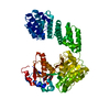
| ||||||||
|---|---|---|---|---|---|---|---|---|---|
| 1 |
| ||||||||
| Unit cell |
|
-Components
| #1: Protein | Mass: 60010.074 Da / Num. of mol.: 1 / Fragment: UNP residues 33-575 Source method: isolated from a genetically manipulated source Details: LpoA residues 33-575 / Source: (gene. exp.) Haemophilus influenzae (bacteria) / Strain: ATCC 51907 / DSM 11121 / KW20 / Rd / Gene: lpoA, HI_1655 / Plasmid: pETBlue-2 / Details (production host): T7 promoter / Production host: |
|---|---|
| #2: Chemical | ChemComp-CL /   Mass: 35.453 Da / Num. of mol.: 1 / Source method: obtained synthetically / Formula: Cl Mass: 35.453 Da / Num. of mol.: 1 / Source method: obtained synthetically / Formula: Cl |
| #3: Water | ChemComp-HOH /  Mass: 18.015 Da / Num. of mol.: 392 / Source method: isolated from a natural source / Formula: H2O Mass: 18.015 Da / Num. of mol.: 392 / Source method: isolated from a natural source / Formula: H2O |
| Has protein modification | Y |
-Experimental details
-Experiment
| Experiment | Method: X-RAY DIFFRACTION / Number of used crystals: 1 |
|---|
- Sample preparation
| Crystal | Density Matthews: 2.4 Å3/Da / Density % sol: 48.79 % |
|---|---|
| Crystal grow | Temperature: 295 K / Method: vapor diffusion, hanging drop / pH: 4 Details: 1 microliter of concentrated protein (33 mg/ml in 150 mM NaCl, 50 mM Tris-HCl, pH 8.0) and 1 microliter precipitant [25% w/v PEG 1500, 0.1 M MMT buffer (20 mM DL-malic acid, 40 mM MES, 40 mM ...Details: 1 microliter of concentrated protein (33 mg/ml in 150 mM NaCl, 50 mM Tris-HCl, pH 8.0) and 1 microliter precipitant [25% w/v PEG 1500, 0.1 M MMT buffer (20 mM DL-malic acid, 40 mM MES, 40 mM Tris-HCl), pH 4.0] and 0.2 microliter 30% xylitol Temp details: 277 for 2 days, then 295 |
-Data collection
| Diffraction | Mean temperature: 125 K | |||||||||||||||||||||||||||||||||||||||||||||||||||||||||||||||||||||||||||||||||||||||||||||||||||||||||
|---|---|---|---|---|---|---|---|---|---|---|---|---|---|---|---|---|---|---|---|---|---|---|---|---|---|---|---|---|---|---|---|---|---|---|---|---|---|---|---|---|---|---|---|---|---|---|---|---|---|---|---|---|---|---|---|---|---|---|---|---|---|---|---|---|---|---|---|---|---|---|---|---|---|---|---|---|---|---|---|---|---|---|---|---|---|---|---|---|---|---|---|---|---|---|---|---|---|---|---|---|---|---|---|---|---|---|
| Diffraction source | Source: SYNCHROTRON / Site: APS / Beamline: 21-ID-G / Wavelength: 0.9786 Å | |||||||||||||||||||||||||||||||||||||||||||||||||||||||||||||||||||||||||||||||||||||||||||||||||||||||||
| Detector | Type: RAYONIX MX-300 / Detector: CCD / Date: Jul 13, 2009 / Details: Monochromator | |||||||||||||||||||||||||||||||||||||||||||||||||||||||||||||||||||||||||||||||||||||||||||||||||||||||||
| Radiation | Monochromator: 0.9786 / Protocol: SINGLE WAVELENGTH / Monochromatic (M) / Laue (L): M / Scattering type: x-ray | |||||||||||||||||||||||||||||||||||||||||||||||||||||||||||||||||||||||||||||||||||||||||||||||||||||||||
| Radiation wavelength | Wavelength: 0.9786 Å / Relative weight: 1 | |||||||||||||||||||||||||||||||||||||||||||||||||||||||||||||||||||||||||||||||||||||||||||||||||||||||||
| Reflection | Resolution: 1.965→50 Å / Num. obs: 42000 / % possible obs: 99.8 % / Redundancy: 6.5 % / Biso Wilson estimate: 31.05 Å2 / Rmerge(I) obs: 0.058 / Net I/av σ(I): 29.518 / Net I/σ(I): 21.6 | |||||||||||||||||||||||||||||||||||||||||||||||||||||||||||||||||||||||||||||||||||||||||||||||||||||||||
| Reflection shell |
|
- Processing
| Software |
| ||||||||||||||||||||||||||||||||||||||||||||||||||||||||||||||||||||||||||||||||||||||||||||||||||||||||||||||||||||||||||||||||||||||||||||||||||||||
|---|---|---|---|---|---|---|---|---|---|---|---|---|---|---|---|---|---|---|---|---|---|---|---|---|---|---|---|---|---|---|---|---|---|---|---|---|---|---|---|---|---|---|---|---|---|---|---|---|---|---|---|---|---|---|---|---|---|---|---|---|---|---|---|---|---|---|---|---|---|---|---|---|---|---|---|---|---|---|---|---|---|---|---|---|---|---|---|---|---|---|---|---|---|---|---|---|---|---|---|---|---|---|---|---|---|---|---|---|---|---|---|---|---|---|---|---|---|---|---|---|---|---|---|---|---|---|---|---|---|---|---|---|---|---|---|---|---|---|---|---|---|---|---|---|---|---|---|---|---|---|---|
| Refinement | Method to determine structure: MOLECULAR REPLACEMENT Starting model: 4P29 and 3CKM Resolution: 1.965→31.783 Å / SU ML: 0.18 / Cross valid method: FREE R-VALUE / σ(F): 1.34 / Phase error: 21.72 / Stereochemistry target values: ML
| ||||||||||||||||||||||||||||||||||||||||||||||||||||||||||||||||||||||||||||||||||||||||||||||||||||||||||||||||||||||||||||||||||||||||||||||||||||||
| Solvent computation | Shrinkage radii: 0.9 Å / VDW probe radii: 1.11 Å / Solvent model: FLAT BULK SOLVENT MODEL | ||||||||||||||||||||||||||||||||||||||||||||||||||||||||||||||||||||||||||||||||||||||||||||||||||||||||||||||||||||||||||||||||||||||||||||||||||||||
| Refinement step | Cycle: LAST / Resolution: 1.965→31.783 Å
| ||||||||||||||||||||||||||||||||||||||||||||||||||||||||||||||||||||||||||||||||||||||||||||||||||||||||||||||||||||||||||||||||||||||||||||||||||||||
| Refine LS restraints |
| ||||||||||||||||||||||||||||||||||||||||||||||||||||||||||||||||||||||||||||||||||||||||||||||||||||||||||||||||||||||||||||||||||||||||||||||||||||||
| LS refinement shell |
| ||||||||||||||||||||||||||||||||||||||||||||||||||||||||||||||||||||||||||||||||||||||||||||||||||||||||||||||||||||||||||||||||||||||||||||||||||||||
| Refinement TLS params. | Method: refined / Refine-ID: X-RAY DIFFRACTION
| ||||||||||||||||||||||||||||||||||||||||||||||||||||||||||||||||||||||||||||||||||||||||||||||||||||||||||||||||||||||||||||||||||||||||||||||||||||||
| Refinement TLS group |
|
