 Movie
Movie Controller

[English] 日本語
 Yorodumi
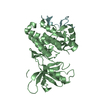
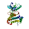
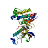
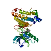
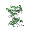
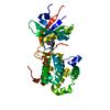
- PDB-4x7h: Co-crystal Structure of PERK bound to N-{5-[(6,7-dimethoxyquinoli... -
+ Open data

- Basic information
| Entry | Database: PDB / ID: 4x7h | ||||||
|---|---|---|---|---|---|---|---|
| Title | Co-crystal Structure of PERK bound to N-{5-[(6,7-dimethoxyquinolin-4-yl)oxy]pyridin-2-yl}-1-methyl-3-oxo-2-phenyl-5-(pyridin-4-yl)-2,3-dihydro-1H-pyrazole-4-carboxamide inhibitor | ||||||
 Components Components | Eukaryotic translation initiation factor 2-alpha kinase 3,Eukaryotic translation initiation factor 2-alpha kinase 3 | ||||||
 Keywords Keywords | TRANSFERASE/TRANSFERASE INHIBITOR /  CATALYTIC DOMAIN / TRANSFERASE-TRANSFERASE INHIBITOR / COMPLEX / TRANSFERASE-TRANSFERASE INHIBITOR complex CATALYTIC DOMAIN / TRANSFERASE-TRANSFERASE INHIBITOR / COMPLEX / TRANSFERASE-TRANSFERASE INHIBITOR complex | ||||||
| Function / homology |  Function and homology information Function and homology informationregulation of endoplasmic reticulum stress-induced eIF2 alpha phosphorylation / negative regulation of translation in response to stress / regulation of endoplasmic reticulum stress-induced intrinsic apoptotic signaling pathway / regulation of translational initiation by eIF2 alpha phosphorylation / eiF2alpha phosphorylation in response to endoplasmic reticulum stress / chondrocyte development / eukaryotic translation initiation factor 2alpha kinase activity / response to manganese-induced endoplasmic reticulum stress / negative regulation of translational initiation in response to stress / PERK-mediated unfolded protein response ...regulation of endoplasmic reticulum stress-induced eIF2 alpha phosphorylation / negative regulation of translation in response to stress / regulation of endoplasmic reticulum stress-induced intrinsic apoptotic signaling pathway / regulation of translational initiation by eIF2 alpha phosphorylation / eiF2alpha phosphorylation in response to endoplasmic reticulum stress / chondrocyte development / eukaryotic translation initiation factor 2alpha kinase activity / response to manganese-induced endoplasmic reticulum stress / negative regulation of translational initiation in response to stress / PERK-mediated unfolded protein response / PERK regulates gene expression / negative regulation of myelination / endocrine pancreas development / ALK mutants bind TKIs / endoplasmic reticulum organization / cellular response to cold / positive regulation of transcription by RNA polymerase I / ER overload response / bone mineralization / Signaling by ALK fusions and activated point mutants / positive regulation of vascular endothelial growth factor production / cellular response to glucose starvation / endoplasmic reticulum unfolded protein response / negative regulation of translational initiation / cellular response to amino acid starvation / response to endoplasmic reticulum stress / ossification / insulin-like growth factor receptor signaling pathway / skeletal system development / calcium-mediated signaling / Hsp90 protein binding / positive regulation of protein localization to nucleus / activation of cysteine-type endopeptidase activity involved in apoptotic process / KEAP1-NFE2L2 pathway / protein phosphatase binding / peptidyl-serine phosphorylation / angiogenesis / negative regulation of translation / protein autophosphorylation / non-specific serine/threonine protein kinase / protein kinase activity / protein phosphorylation / protein serine kinase activity / protein serine/threonine kinase activity / endoplasmic reticulum membrane / positive regulation of gene expression / perinuclear region of cytoplasm / enzyme binding / endoplasmic reticulum / ATP binding / membrane / identical protein binding / nucleus / cytosol / cytoplasmSimilarity search - Function | ||||||
| Biological species |  Homo sapiens (human) Homo sapiens (human) | ||||||
| Method | X-RAY DIFFRACTION / SYNCHROTRON / MOLECULAR REPLACEMENT / Resolution: 2 Å | ||||||
 Authors Authors | Shaffer, P.L. / Bellon, S.F. / Long, A.M. / Chen, H. | ||||||
 Citation Citation | Journal: J.Med.Chem. / Year: 2015 Title: Discovery of 1H-Pyrazol-3(2H)-ones as Potent and Selective Inhibitors of Protein Kinase R-like Endoplasmic Reticulum Kinase (PERK). Authors: Smith, A.L. / Andrews, K.L. / Beckmann, H. / Bellon, S.F. / Beltran, P.J. / Booker, S. / Chen, H. / Chung, Y.A. / D'Angelo, N.D. / Dao, J. / Dellamaggiore, K.R. / Jaeckel, P. / Kendall, R. / ...Authors: Smith, A.L. / Andrews, K.L. / Beckmann, H. / Bellon, S.F. / Beltran, P.J. / Booker, S. / Chen, H. / Chung, Y.A. / D'Angelo, N.D. / Dao, J. / Dellamaggiore, K.R. / Jaeckel, P. / Kendall, R. / Labitzke, K. / Long, A.M. / Materna-Reichelt, S. / Mitchell, P. / Norman, M.H. / Powers, D. / Rose, M. / Shaffer, P.L. / Wu, M.M. / Lipford, J.R. | ||||||
| History |
|
- Structure visualization
| Structure viewer | Molecule: MolmilJmol/JSmol |
|---|
- Downloads & links
-Download
| PDBx/mmCIF format | 4x7h.cif.gz | 130 KB | Display | PDBx/mmCIF format |
|---|---|---|---|---|
| PDB format | pdb4x7h.ent.gz | 98.5 KB | Display | PDB format |
| PDBx/mmJSON format | 4x7h.json.gz | Tree view | PDBx/mmJSON format | |
| Others |  Other downloads Other downloads |
-Validation report
| Arichive directory | https://data.pdbj.org/pub/pdb/validation_reports/x7/4x7hftp://data.pdbj.org/pub/pdb/validation_reports/x7/4x7h | HTTPS FTP |
|---|
-Related structure data
| Related structure data |  4x7jC  4x7kC  4x7lC  4x7nC  4x7oC 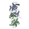 1zy4S C: citing same article ( S: Starting model for refinement |
|---|---|
| Similar structure data |


-Links
 PDBj
PDBj- Assembly
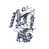
| Deposited unit | 
| ||||||||
|---|---|---|---|---|---|---|---|---|---|
| 1 |
| ||||||||
| Unit cell |
| ||||||||
| Components on special symmetry positions |
|
-Components
| #1: Protein | Mass: 36816.363 Da / Num. of mol.: 1 / Mutation: D937N, deletion of 670-874 Source method: isolated from a genetically manipulated source Source: (gene. exp.) Homo sapiens (human) / Gene: EIF2AK3, PEK, PERK / Production host:  Escherichia coli (E. coli) Escherichia coli (E. coli)References: UniProt: Q9NZJ5, non-specific serine/threonine protein kinase | ||
|---|---|---|---|
| #2: Chemical | ChemComp-3Z2 / 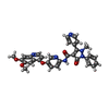  Mass: 574.586 Da / Num. of mol.: 1 / Source method: obtained synthetically / Formula: C32H26N6O5 Mass: 574.586 Da / Num. of mol.: 1 / Source method: obtained synthetically / Formula: C32H26N6O5 | ||
| #3: Chemical | Sulfate  Mass: 96.063 Da / Num. of mol.: 2 / Source method: obtained synthetically / Formula: SO4 Mass: 96.063 Da / Num. of mol.: 2 / Source method: obtained synthetically / Formula: SO4#4: Water | ChemComp-HOH / | Water Mass: 18.015 Da / Num. of mol.: 183 / Source method: isolated from a natural source / Formula: H2O Mass: 18.015 Da / Num. of mol.: 183 / Source method: isolated from a natural source / Formula: H2O |
-Experimental details
-Experiment
| Experiment | Method: X-RAY DIFFRACTION / Number of used crystals: 1 |
|---|
- Sample preparation
| Crystal | Density Matthews: 2.88 Å3/Da / Density % sol: 57.29 % |
|---|---|
| Crystal grow | Temperature: 277 K / Method: vapor diffusion, hanging drop / pH: 6.5 / Details: 14% PEG 3350, 0.3M Sodium Sulfate, 0.1M BTP pH 6.5 |
-Data collection
| Diffraction | Mean temperature: 80 K | ||||||||||||||||||||||||||||||||||||||||||||||||||||||||||||||||||
|---|---|---|---|---|---|---|---|---|---|---|---|---|---|---|---|---|---|---|---|---|---|---|---|---|---|---|---|---|---|---|---|---|---|---|---|---|---|---|---|---|---|---|---|---|---|---|---|---|---|---|---|---|---|---|---|---|---|---|---|---|---|---|---|---|---|---|---|
| Diffraction source | Source: SYNCHROTRON / Site: ALS  / Beamline: 5.0.2 / Wavelength: 1 Å / Beamline: 5.0.2 / Wavelength: 1 Å | ||||||||||||||||||||||||||||||||||||||||||||||||||||||||||||||||||
| Detector | Type: ADSC QUANTUM 315 / Detector: CCD / Date: Aug 18, 2008 | ||||||||||||||||||||||||||||||||||||||||||||||||||||||||||||||||||
| Radiation | Protocol: SINGLE WAVELENGTH / Monochromatic (M) / Laue (L): M / Scattering type: x-ray | ||||||||||||||||||||||||||||||||||||||||||||||||||||||||||||||||||
| Radiation wavelength | Wavelength: 1 Å / Relative weight: 1 | ||||||||||||||||||||||||||||||||||||||||||||||||||||||||||||||||||
| Reflection | Resolution: 2→50 Å / Num. obs: 29688 / % possible obs: 100 % / Redundancy: 13.8 % / Rmerge(I) obs: 0.097 / Χ2: 1.131 / Net I/av σ(I): 26.938 / Net I/σ(I): 12.3 / Num. measured all: 410972 | ||||||||||||||||||||||||||||||||||||||||||||||||||||||||||||||||||
| Reflection shell | Diffraction-ID: 1 / Rejects: 0
|
- Processing
| Software |
| |||||||||||||||||||||||||||||||||||||||||||||||||||||||||||||||||||||||||||
|---|---|---|---|---|---|---|---|---|---|---|---|---|---|---|---|---|---|---|---|---|---|---|---|---|---|---|---|---|---|---|---|---|---|---|---|---|---|---|---|---|---|---|---|---|---|---|---|---|---|---|---|---|---|---|---|---|---|---|---|---|---|---|---|---|---|---|---|---|---|---|---|---|---|---|---|---|
| Refinement | Method to determine structure: MOLECULAR REPLACEMENT Starting model: 1ZY4 Resolution: 2→37.81 Å / Cor.coef. Fo:Fc: 0.959 / Cor.coef. Fo:Fc free: 0.957 / SU B: 5.987 / SU ML: 0.088 / Cross valid method: THROUGHOUT / σ(F): 0 / ESU R: 0.147 / ESU R Free: 0.127 / Stereochemistry target values: MAXIMUM LIKELIHOOD Details: HYDROGENS HAVE BEEN USED IF PRESENT IN THE INPUT U VALUES : WITH TLS ADDED
| |||||||||||||||||||||||||||||||||||||||||||||||||||||||||||||||||||||||||||
| Solvent computation | Ion probe radii: 0.8 Å / Shrinkage radii: 0.8 Å / VDW probe radii: 1.4 Å / Solvent model: MASK | |||||||||||||||||||||||||||||||||||||||||||||||||||||||||||||||||||||||||||
| Displacement parameters | Biso max: 88.54 Å2 / Biso mean: 45.115 Å2 / Biso min: 25.67 Å2
| |||||||||||||||||||||||||||||||||||||||||||||||||||||||||||||||||||||||||||
| Refinement step | Cycle: final / Resolution: 2→37.81 Å
| |||||||||||||||||||||||||||||||||||||||||||||||||||||||||||||||||||||||||||
| Refine LS restraints |
| |||||||||||||||||||||||||||||||||||||||||||||||||||||||||||||||||||||||||||
| LS refinement shell | Resolution: 2.003→2.055 Å / Total num. of bins used: 20
| |||||||||||||||||||||||||||||||||||||||||||||||||||||||||||||||||||||||||||
| Refinement TLS params. | Method: refined / Refine-ID: X-RAY DIFFRACTION
| |||||||||||||||||||||||||||||||||||||||||||||||||||||||||||||||||||||||||||
| Refinement TLS group |
|
