 Movie
Movie Controller

[English] 日本語
 Yorodumi
- PDB-1zy4: Crystal Structure of eIF2alpha Protein Kinase GCN2: R794G Hyperac... -
+ Open data

- Basic information
| Entry | Database: PDB / ID: 1zy4 | ||||||
|---|---|---|---|---|---|---|---|
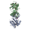
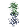
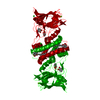
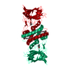
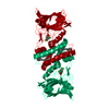
| Title | Crystal Structure of eIF2alpha Protein Kinase GCN2: R794G Hyperactivating Mutant in Apo Form. | ||||||
 Components Components | Serine/threonine-protein kinase GCN2 | ||||||
 Keywords Keywords | TRANSFERASE / TRANSLATION REGULATOR / PROTEIN KINASE / SIGNAL TRANSDUCTION / AMINO-ACID STARVATION / STARVATION STRESS RESPONSE / EIF2ALPHA KINASE | ||||||
| Function / homology |  Function and homology information Function and homology informationcellular response to histidine / regulation of cytoplasmic translational initiation in response to stress / positive regulation of translational initiation in response to starvation / GCN2-mediated signaling / eukaryotic translation initiation factor 2alpha kinase activity / negative regulation of translational initiation in response to stress / positive regulation of cellular response to amino acid starvation / regulation of translational initiation / ribosomal large subunit binding / protein kinase inhibitor activity ...cellular response to histidine / regulation of cytoplasmic translational initiation in response to stress / positive regulation of translational initiation in response to starvation / GCN2-mediated signaling / eukaryotic translation initiation factor 2alpha kinase activity / negative regulation of translational initiation in response to stress / positive regulation of cellular response to amino acid starvation / regulation of translational initiation / ribosomal large subunit binding / protein kinase inhibitor activity / DNA damage checkpoint signaling / translation initiation factor binding / cellular response to amino acid starvation / cytosolic ribosome / protein autophosphorylation / double-stranded RNA binding / large ribosomal subunit / ribosome binding / small ribosomal subunit / protein phosphorylation / protein kinase activity / non-specific serine/threonine protein kinase / tRNA binding / intracellular signal transduction / translation / protein serine kinase activity / protein homodimerization activity / ATP binding / nucleus / cytosol / cytoplasm Similarity search - Function | ||||||
| Biological species |  | ||||||
| Method |  X-RAY DIFFRACTION / SYNCHROTRON / MOLECULAR REPLACEMENT / Resolution: 1.95 Å X-RAY DIFFRACTION / SYNCHROTRON / MOLECULAR REPLACEMENT / Resolution: 1.95 Å | ||||||
 Authors Authors | Padyana, A.K. / Qiu, H. / Roll-Mecak, A. / Hinnebusch, A.G. / Burley, S.K. | ||||||
 Citation Citation | Journal: J.Biol.Chem. / Year: 2005 Title: Structural Basis for Autoinhibition and Mutational Activation of Eukaryotic Initiation Factor 2{alpha} Protein Kinase GCN2 Authors: Padyana, A.K. / Qiu, H. / Roll-Mecak, A. / Hinnebusch, A.G. / Burley, S.K. #1: Journal: Genes Dev. / Year: 2002Title: Mutations that Bypass tRNA Binding Activate the Intrinsically Defective Kinase Domain in GCN2. Authors: Qiu, H. / Hu, C. / Dong, J. / Hinnebusch, A.G. | ||||||
| History |
| ||||||
| Remark 600 | HETEROGEN GOL 997 is associated with protein chain A. GOL 998 is associated with protein chain B. |
- Structure visualization
| Structure viewer | Molecule: MolmilJmol/JSmol |
|---|
- Downloads & links
-Download
| PDBx/mmCIF format | 1zy4.cif.gz | 125.1 KB | Display | PDBx/mmCIF format |
|---|---|---|---|---|
| PDB format | pdb1zy4.ent.gz | 96.8 KB | Display | PDB format |
| PDBx/mmJSON format | 1zy4.json.gz | Tree view | PDBx/mmJSON format | |
| Others |  Other downloads Other downloads |
-Validation report
| Arichive directory | https://data.pdbj.org/pub/pdb/validation_reports/zy/1zy4ftp://data.pdbj.org/pub/pdb/validation_reports/zy/1zy4 | HTTPS FTP |
|---|
-Related structure data
| Related structure data |  1zxeC  1zy5C  1zycSC 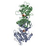 1zydC C: citing same article ( S: Starting model for refinement |
|---|---|
| Similar structure data |

-Links
 PDBj
PDBj


- Assembly
| Deposited unit | 
| ||||||||
|---|---|---|---|---|---|---|---|---|---|
| 1 |
| ||||||||
| Unit cell |
| ||||||||
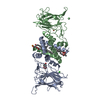
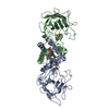
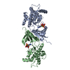
| Details | The biological assembly of GCN2 protein kinase is a dimer. The asymmetric unit of this crystal lattice contains one dimer assembled into one biological unit. |
-Components
| #1: Protein | Mass: 35043.336 Da / Num. of mol.: 2 / Mutation: R794G Source method: isolated from a genetically manipulated source Source: (gene. exp.) Gene: GCN2, AAS1 / Plasmid: pET26b / Production host:  #2: Chemical |   Mass: 92.094 Da / Num. of mol.: 2 / Source method: obtained synthetically / Formula: C3H8O3 Mass: 92.094 Da / Num. of mol.: 2 / Source method: obtained synthetically / Formula: C3H8O3#3: Water | ChemComp-HOH / |  Mass: 18.015 Da / Num. of mol.: 234 / Source method: isolated from a natural source / Formula: H2O Mass: 18.015 Da / Num. of mol.: 234 / Source method: isolated from a natural source / Formula: H2O |
|---|
-Experimental details
-Experiment
| Experiment | Method: X-RAY DIFFRACTION / Number of used crystals: 1 |
|---|
- Sample preparation
| Crystal | Density Matthews: 2.2 Å3/Da / Density % sol: 43.97 % |
|---|---|
| Crystal grow | Temperature: 295 K / Method: vapor diffusion, sitting drop / pH: 9.5 Details: PEG 6000, CHES, pH 9.5, VAPOR DIFFUSION, SITTING DROP, temperature 295K |
-Data collection
| Diffraction | Mean temperature: 100 K |
|---|---|
| Diffraction source | Source: SYNCHROTRON / Site: APS  / Beamline: 31-ID / Wavelength: 0.97931 Å / Beamline: 31-ID / Wavelength: 0.97931 Å |
| Detector | Type: MARRESEARCH / Detector: CCD / Date: Aug 19, 2004 |
| Radiation | Monochromator: DIAMOND 111 / Protocol: SINGLE WAVELENGTH / Monochromatic (M) / Laue (L): M / Scattering type: x-ray |
| Radiation wavelength | Wavelength: 0.97931 Å / Relative weight: 1 |
| Reflection | Resolution: 1.95→50 Å / Num. all: 45568 / Num. obs: 45176 / % possible obs: 99.1 % / Observed criterion σ(F): 0 / Observed criterion σ(I): 0 / Redundancy: 9.9 % / Biso Wilson estimate: 25.4 Å2 / Rmerge(I) obs: 0.068 / Net I/σ(I): 22.4 |
| Reflection shell | Resolution: 1.95→2 Å / Rmerge(I) obs: 0.66 / Mean I/σ(I) obs: 2.8 / % possible all: 97.6 |
- Processing
| Software |
| ||||||||||||||||||||||||||||||||||||||||||||||||||||||||||||||||||||||||||||||||
|---|---|---|---|---|---|---|---|---|---|---|---|---|---|---|---|---|---|---|---|---|---|---|---|---|---|---|---|---|---|---|---|---|---|---|---|---|---|---|---|---|---|---|---|---|---|---|---|---|---|---|---|---|---|---|---|---|---|---|---|---|---|---|---|---|---|---|---|---|---|---|---|---|---|---|---|---|---|---|---|---|---|
| Refinement | Method to determine structure: MOLECULAR REPLACEMENT Starting model: 1ZYC Resolution: 1.95→37.72 Å / Rfactor Rfree error: 0.007 / Data cutoff high absF: 1973272.77 / Data cutoff low absF: 0 / Isotropic thermal model: RESTRAINED / Cross valid method: THROUGHOUT / σ(F): 0 / Stereochemistry target values: Engh & Huber
| ||||||||||||||||||||||||||||||||||||||||||||||||||||||||||||||||||||||||||||||||
| Solvent computation | Solvent model: FLAT MODEL / Bsol: 60.6746 Å2 / ksol: 0.359013 e/Å3 | ||||||||||||||||||||||||||||||||||||||||||||||||||||||||||||||||||||||||||||||||
| Displacement parameters | Biso mean: 48.6 Å2
| ||||||||||||||||||||||||||||||||||||||||||||||||||||||||||||||||||||||||||||||||
| Refine analyze |
| ||||||||||||||||||||||||||||||||||||||||||||||||||||||||||||||||||||||||||||||||
| Refinement step | Cycle: LAST / Resolution: 1.95→37.72 Å
| ||||||||||||||||||||||||||||||||||||||||||||||||||||||||||||||||||||||||||||||||
| Refine LS restraints |
| ||||||||||||||||||||||||||||||||||||||||||||||||||||||||||||||||||||||||||||||||
| LS refinement shell | Resolution: 1.95→2.07 Å / Rfactor Rfree error: 0.022 / Total num. of bins used: 6
| ||||||||||||||||||||||||||||||||||||||||||||||||||||||||||||||||||||||||||||||||
| Xplor file |
|
