 Movie
Movie Controller

[English] 日本語
 Yorodumi
- PDB-4u9b: Crystal structure of an H-NOX protein from S. oneidensis in the F... -
+ Open data

- Basic information
| Entry | Database: PDB / ID: 4u9b | ||||||
|---|---|---|---|---|---|---|---|
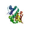
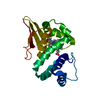
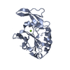
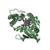
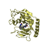
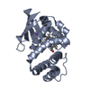
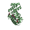
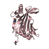
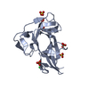
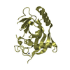
| Title | Crystal structure of an H-NOX protein from S. oneidensis in the Fe(II)NO ligation state | ||||||
 Components Components | NO-binding heme-dependent sensor protein | ||||||
 Keywords Keywords | SIGNALING PROTEIN / H-NOX / hemoprotein / gas sensor | ||||||
| Function / homology |  Function and homology information Function and homology information | ||||||
| Biological species |  Shewanella oneidensis (bacteria) Shewanella oneidensis (bacteria) | ||||||
| Method |  X-RAY DIFFRACTION / SYNCHROTRON / MOLECULAR REPLACEMENT / Resolution: 1.65 Å X-RAY DIFFRACTION / SYNCHROTRON / MOLECULAR REPLACEMENT / Resolution: 1.65 Å | ||||||
 Authors Authors | Herzik Jr., M.A. / Jonnalagadda, R. / Kuriyan, J. / Marletta, M.A. | ||||||
| Funding support |  United States, 1items United States, 1items
| ||||||
 Citation Citation | Journal: Proc.Natl.Acad.Sci.USA / Year: 2014 Title: Structural insights into the role of iron-histidine bond cleavage in nitric oxide-induced activation of H-NOX gas sensor proteins. Authors: Herzik, M.A. / Jonnalagadda, R. / Kuriyan, J. / Marletta, M.A. | ||||||
| History |
|
- Structure visualization
| Structure viewer | Molecule: MolmilJmol/JSmol |
|---|
- Downloads & links
-Download
| PDBx/mmCIF format | 4u9b.cif.gz | 126.2 KB | Display | PDBx/mmCIF format |
|---|---|---|---|---|
| PDB format | pdb4u9b.ent.gz | 98.9 KB | Display | PDB format |
| PDBx/mmJSON format | 4u9b.json.gz | Tree view | PDBx/mmJSON format | |
| Others |  Other downloads Other downloads |
-Validation report
| Arichive directory | https://data.pdbj.org/pub/pdb/validation_reports/u9/4u9bftp://data.pdbj.org/pub/pdb/validation_reports/u9/4u9b | HTTPS FTP |
|---|
-Related structure data
| Related structure data | 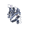 4u99SC 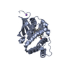 4u9gC  4u9jC 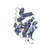 4u9kC S: Starting model for refinement C: citing same article ( |
|---|---|
| Similar structure data |


-Links
 PDBj
PDBj


- Assembly
| Deposited unit | 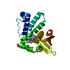
| ||||||||
|---|---|---|---|---|---|---|---|---|---|
| 1 |
| ||||||||
| Unit cell |
|
-Components
-Protein , 1 types, 1 molecules A
| #1: Protein | Mass: 21329.447 Da / Num. of mol.: 1 Source method: isolated from a genetically manipulated source Source: (gene. exp.) Shewanella oneidensis (bacteria) / Strain: MR-1 / Gene: SO_2144 / Plasmid: pET20 / Production host: |
|---|
-Non-polymers , 6 types, 126 molecules 


| #2: Chemical | ChemComp-HEM /  Mass: 616.487 Da / Num. of mol.: 1 / Source method: obtained synthetically / Formula: C34H32FeN4O4 Mass: 616.487 Da / Num. of mol.: 1 / Source method: obtained synthetically / Formula: C34H32FeN4O4 |
|---|---|
| #3: Chemical | ChemComp-NO /  Mass: 30.006 Da / Num. of mol.: 1 / Source method: obtained synthetically / Formula: NO Mass: 30.006 Da / Num. of mol.: 1 / Source method: obtained synthetically / Formula: NO |
| #4: Chemical | ChemComp-ZN /  Mass: 65.409 Da / Num. of mol.: 1 / Source method: obtained synthetically / Formula: Zn Mass: 65.409 Da / Num. of mol.: 1 / Source method: obtained synthetically / Formula: Zn |
| #5: Chemical | ChemComp-PO4 /  Mass: 94.971 Da / Num. of mol.: 1 / Source method: obtained synthetically / Formula: PO4 Mass: 94.971 Da / Num. of mol.: 1 / Source method: obtained synthetically / Formula: PO4 |
| #6: Chemical | ChemComp-GOL /  Mass: 92.094 Da / Num. of mol.: 1 / Source method: obtained synthetically / Formula: C3H8O3 Mass: 92.094 Da / Num. of mol.: 1 / Source method: obtained synthetically / Formula: C3H8O3 |
| #7: Water | ChemComp-HOH / Mass: 18.015 Da / Num. of mol.: 121 / Source method: isolated from a natural source / Formula: H2O |
-Experimental details
-Experiment
| Experiment | Method: X-RAY DIFFRACTION / Number of used crystals: 1 |
|---|
- Sample preparation
| Crystal | Density Matthews: 2.36 Å3/Da / Density % sol: 47.84 % |
|---|---|
| Crystal grow | Temperature: 293 K / Method: vapor diffusion, sitting drop Details: Obtained by equilibrating a 2 or 4 uL drop of 1:1 protein: reservoir against 700 uL reservoir containing 0.7 M NaH2PO4, 0.9 M K2HPO4. Cryoprotection was achieved by carefully adding equal ...Details: Obtained by equilibrating a 2 or 4 uL drop of 1:1 protein: reservoir against 700 uL reservoir containing 0.7 M NaH2PO4, 0.9 M K2HPO4. Cryoprotection was achieved by carefully adding equal drop volume of mother liquor containing 50% glycerol. Crystals were then transferred into mother liquor containing 30% glycerol and flash frozen in liquid nitrogen. All crystal growth and manipulation was performed anaerobically. |
-Data collection
| Diffraction | Mean temperature: 100 K |
|---|---|
| Diffraction source | Source: SYNCHROTRON / Site: ALS / Beamline: 8.2.1 / Wavelength: 1.11 Å |
| Detector | Type: ADSC QUANTUM 315r / Detector: CCD / Date: Jul 7, 2012 |
| Radiation | Protocol: SINGLE WAVELENGTH / Monochromatic (M) / Laue (L): M / Scattering type: x-ray |
| Radiation wavelength | Wavelength: 1.11 Å / Relative weight: 1 |
| Reflection | Resolution: 1.65→33.818 Å / Num. all: 45550 / Num. obs: 45550 / % possible obs: 98.46 % / Redundancy: 3.9 % / Rmerge(I) obs: 0.029 / Net I/σ(I): 14.4 |
| Reflection shell | Resolution: 1.65→1.74 Å / Redundancy: 3.3 % / Rmerge(I) obs: 0.487 / Mean I/σ(I) obs: 1.7 / % possible all: 99.5 |
- Processing
| Software | Name: PHENIX / Version: (phenix.refine: 1.9_1692) / Classification: refinement | |||||||||||||||||||||||||||||||||||||||||||||||||||||||||||||||||||||||||||||||||||||||||||||||||||||||||||||||||||||||
|---|---|---|---|---|---|---|---|---|---|---|---|---|---|---|---|---|---|---|---|---|---|---|---|---|---|---|---|---|---|---|---|---|---|---|---|---|---|---|---|---|---|---|---|---|---|---|---|---|---|---|---|---|---|---|---|---|---|---|---|---|---|---|---|---|---|---|---|---|---|---|---|---|---|---|---|---|---|---|---|---|---|---|---|---|---|---|---|---|---|---|---|---|---|---|---|---|---|---|---|---|---|---|---|---|---|---|---|---|---|---|---|---|---|---|---|---|---|---|---|---|
| Refinement | Method to determine structure: MOLECULAR REPLACEMENT Starting model: 4U99 Resolution: 1.65→33.818 Å / SU ML: 0.21 / Cross valid method: FREE R-VALUE / σ(F): 1.33 / Phase error: 22.91 / Stereochemistry target values: ML
| |||||||||||||||||||||||||||||||||||||||||||||||||||||||||||||||||||||||||||||||||||||||||||||||||||||||||||||||||||||||
| Solvent computation | Shrinkage radii: 0.6 Å / VDW probe radii: 1 Å / Solvent model: FLAT BULK SOLVENT MODEL | |||||||||||||||||||||||||||||||||||||||||||||||||||||||||||||||||||||||||||||||||||||||||||||||||||||||||||||||||||||||
| Refinement step | Cycle: LAST / Resolution: 1.65→33.818 Å
| |||||||||||||||||||||||||||||||||||||||||||||||||||||||||||||||||||||||||||||||||||||||||||||||||||||||||||||||||||||||
| Refine LS restraints |
| |||||||||||||||||||||||||||||||||||||||||||||||||||||||||||||||||||||||||||||||||||||||||||||||||||||||||||||||||||||||
| LS refinement shell |
|
