 Movie
Movie Controller

[English] 日本語
 Yorodumi
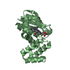
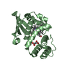
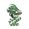
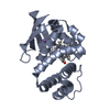
- PDB-7lgk: Crystal structure of soluble guanylate cyclase activator runcacig... -
+ Open data

- Basic information
| Entry | Database: PDB / ID: 7lgk | ||||||
|---|---|---|---|---|---|---|---|
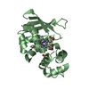
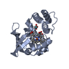
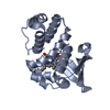
| Title | Crystal structure of soluble guanylate cyclase activator runcaciguat (BAY 1101042) bound to nostoc H-NOX domain | ||||||
 Components Components | H-NOX domain protein | ||||||
 Keywords Keywords | SIGNALING PROTEIN/ACTIVATOR / Soluble guanylyl cyclase / H-NOX / signal transduction / activator / SIGNALING PROTEIN / SIGNALING PROTEIN-SIGNALING PROTEIN ACTIVATOR complex / SIGNALING PROTEIN-ACTIVATOR complex | ||||||
| Function / homology | Heme NO-binding / H-NOX domain superfamily / Haem-NO-binding / NO signalling/Golgi transport ligand-binding domain superfamily / heme binding / metal ion binding / Runcaciguat / Alr2278 protein Function and homology information Function and homology information | ||||||
| Biological species |  Nostoc sp. PCC 7120 = FACHB-418 (bacteria) Nostoc sp. PCC 7120 = FACHB-418 (bacteria) | ||||||
| Method |  X-RAY DIFFRACTION / SYNCHROTRON / MOLECULAR REPLACEMENT / Resolution: 2.2 Å X-RAY DIFFRACTION / SYNCHROTRON / MOLECULAR REPLACEMENT / Resolution: 2.2 Å | ||||||
 Authors Authors | van den Akker, F. / Kumar, V. / Schaefer, M. | ||||||
 Citation Citation | Journal: J.Med.Chem. / Year: 2021 Title: Discovery of the Soluble Guanylate Cyclase Activator Runcaciguat (BAY 1101042). Authors: Hahn, M.G. / Lampe, T. / El Sheikh, S. / Griebenow, N. / Woltering, E. / Schlemmer, K.H. / Dietz, L. / Gerisch, M. / Wunder, F. / Becker-Pelster, E.M. / Mondritzki, T. / Tinel, H. / Knorr, A. ...Authors: Hahn, M.G. / Lampe, T. / El Sheikh, S. / Griebenow, N. / Woltering, E. / Schlemmer, K.H. / Dietz, L. / Gerisch, M. / Wunder, F. / Becker-Pelster, E.M. / Mondritzki, T. / Tinel, H. / Knorr, A. / Kern, A. / Lang, D. / Hueser, J. / Schomber, T. / Benardeau, A. / Eitner, F. / Truebel, H. / Mittendorf, J. / Kumar, V. / van den Akker, F. / Schaefer, M. / Geiss, V. / Sandner, P. / Stasch, J.P. | ||||||
| History |
|
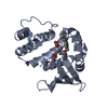
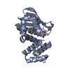
- Structure visualization
| Structure viewer | Molecule: MolmilJmol/JSmol |
|---|
- Downloads & links
-Download
| PDBx/mmCIF format | 7lgk.cif.gz | 164 KB | Display | PDBx/mmCIF format |
|---|---|---|---|---|
| PDB format | pdb7lgk.ent.gz | 128.1 KB | Display | PDB format |
| PDBx/mmJSON format | 7lgk.json.gz | Tree view | PDBx/mmJSON format | |
| Others |  Other downloads Other downloads |
-Validation report
| Arichive directory | https://data.pdbj.org/pub/pdb/validation_reports/lg/7lgkftp://data.pdbj.org/pub/pdb/validation_reports/lg/7lgk | HTTPS FTP |
|---|
-Related structure data
| Related structure data | 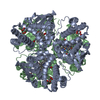 4iamS S: Starting model for refinement |
|---|---|
| Similar structure data |

-Links
 PDBj
PDBj
- Assembly
| Deposited unit | 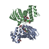
| ||||||||
|---|---|---|---|---|---|---|---|---|---|
| 1 | 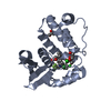
| ||||||||
| 2 | 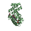
| ||||||||
| Unit cell |
|
-Components
| #1: Protein | Mass: 20358.799 Da / Num. of mol.: 2 Source method: isolated from a genetically manipulated source Source: (gene. exp.) Nostoc sp. PCC 7120 = FACHB-418 (bacteria)Strain: PCC 7120 / SAG 25.82 / UTEX 2576 / Gene: alr2278 / Production host: #2: Chemical |   Mass: 488.327 Da / Num. of mol.: 2 / Source method: obtained synthetically / Formula: C23H22Cl2F3NO3 / Feature type: SUBJECT OF INVESTIGATION Mass: 488.327 Da / Num. of mol.: 2 / Source method: obtained synthetically / Formula: C23H22Cl2F3NO3 / Feature type: SUBJECT OF INVESTIGATION#3: Chemical |   Mass: 35.453 Da / Num. of mol.: 2 / Source method: obtained synthetically / Formula: Cl Mass: 35.453 Da / Num. of mol.: 2 / Source method: obtained synthetically / Formula: Cl#4: Chemical | ChemComp-GOL /   Mass: 92.094 Da / Num. of mol.: 6 / Source method: obtained synthetically / Formula: C3H8O3 Mass: 92.094 Da / Num. of mol.: 6 / Source method: obtained synthetically / Formula: C3H8O3#5: Water | ChemComp-HOH / |  Mass: 18.015 Da / Num. of mol.: 90 / Source method: isolated from a natural source / Formula: H2O Mass: 18.015 Da / Num. of mol.: 90 / Source method: isolated from a natural source / Formula: H2OHas ligand of interest | Y | |
|---|
-Experimental details
-Experiment
| Experiment | Method: X-RAY DIFFRACTION / Number of used crystals: 1 |
|---|
- Sample preparation
| Crystal | Density Matthews: 3.9 Å3/Da / Density % sol: 68.47 % |
|---|---|
| Crystal grow | Temperature: 293 K / Method: vapor diffusion, sitting drop Details: 0.1 M Tris pH 8.5, 12% glycerol, and 1.5 M ammonium sulfate |
-Data collection
| Diffraction | Mean temperature: 100 K / Serial crystal experiment: N |
|---|---|
| Diffraction source | Source: SYNCHROTRON / Site: PETRA III, DESY  / Beamline: P11 / Wavelength: 1.0332 Å / Beamline: P11 / Wavelength: 1.0332 Å |
| Detector | Type: DECTRIS PILATUS3 S 6M / Detector: PIXEL / Date: Sep 14, 2016 |
| Radiation | Protocol: SINGLE WAVELENGTH / Monochromatic (M) / Laue (L): M / Scattering type: x-ray |
| Radiation wavelength | Wavelength: 1.0332 Å / Relative weight: 1 |
| Reflection | Resolution: 2.2→43.84 Å / Num. obs: 32542 / % possible obs: 100 % / Redundancy: 20 % / Rrim(I) all: 0.14 / Net I/σ(I): 18.98 |
| Reflection shell | Resolution: 2.2→2.33 Å / Num. unique obs: 3000 / Rrim(I) all: 1.97 |
- Processing
| Software |
| |||||||||||||||||||||||||||||||||||||||||||||||||||||||||||||||||||||||||||
|---|---|---|---|---|---|---|---|---|---|---|---|---|---|---|---|---|---|---|---|---|---|---|---|---|---|---|---|---|---|---|---|---|---|---|---|---|---|---|---|---|---|---|---|---|---|---|---|---|---|---|---|---|---|---|---|---|---|---|---|---|---|---|---|---|---|---|---|---|---|---|---|---|---|---|---|---|
| Refinement | Method to determine structure: MOLECULAR REPLACEMENT Starting model: 4IAM Resolution: 2.2→43.84 Å / Cor.coef. Fo:Fc: 0.966 / Cor.coef. Fo:Fc free: 0.947 / SU B: 7.812 / SU ML: 0.099 / Cross valid method: THROUGHOUT / σ(F): 0 / ESU R: 0.147 / ESU R Free: 0.146 / Stereochemistry target values: MAXIMUM LIKELIHOOD Details: HYDROGENS HAVE BEEN ADDED IN THE RIDING POSITIONS U VALUES : WITH TLS ADDED
| |||||||||||||||||||||||||||||||||||||||||||||||||||||||||||||||||||||||||||
| Solvent computation | Ion probe radii: 0.8 Å / Shrinkage radii: 0.8 Å / VDW probe radii: 1.2 Å / Solvent model: BABINET MODEL WITH MASK | |||||||||||||||||||||||||||||||||||||||||||||||||||||||||||||||||||||||||||
| Displacement parameters | Biso max: 137.05 Å2 / Biso mean: 58.827 Å2 / Biso min: 36.63 Å2
| |||||||||||||||||||||||||||||||||||||||||||||||||||||||||||||||||||||||||||
| Refinement step | Cycle: final / Resolution: 2.2→43.84 Å
| |||||||||||||||||||||||||||||||||||||||||||||||||||||||||||||||||||||||||||
| Refine LS restraints |
| |||||||||||||||||||||||||||||||||||||||||||||||||||||||||||||||||||||||||||
| LS refinement shell | Resolution: 2.2→2.256 Å / Rfactor Rfree error: 0
| |||||||||||||||||||||||||||||||||||||||||||||||||||||||||||||||||||||||||||
| Refinement TLS params. | Method: refined / Refine-ID: X-RAY DIFFRACTION
| |||||||||||||||||||||||||||||||||||||||||||||||||||||||||||||||||||||||||||
| Refinement TLS group |
|
