解像度: 2.05→86.2 Å / Cor.coef. Fo:Fc: 0.966 / Cor.coef. Fo:Fc free: 0.957 / SU B: 1.359 / SU ML: 0.036 / 交差検証法: THROUGHOUT / σ(F): 1.72 / ESU R: 0.026 / ESU R Free: 0.025 / 立体化学のターゲット値: MAXIMUM LIKELIHOOD / 詳細: HYDROGENS HAVE BEEN ADDED IN THE RIDING POSITIONS
Rfactor
反射数
%反射
Selection details
Rfree
0.19526
1850
4.9 %
RANDOM
Rwork
0.16341
-
-
-
all
0.165
36315
-
-
obs
0.16494
36251
99.82 %
-
溶媒の処理
イオンプローブ半径: 0.8 Å / 減衰半径: 0.8 Å / VDWプローブ半径: 1.4 Å / 溶媒モデル: MASK
 ムービー
ムービー コントローラー


 データを開く
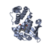
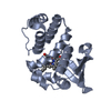
 基本情報
基本情報 要素
要素 キーワード
キーワード 機能・相同性情報
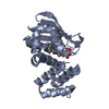
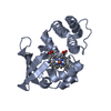
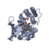
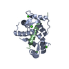
 Nostoc sp. (バクテリア)
 X線回折 /
  データ登録者
 引用
引用 構造の表示
構造の表示 ダウンロードとリンク
 その他のダウンロード
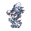


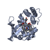
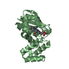
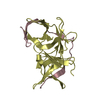
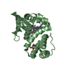
 PDBj
PDBj
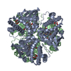
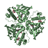
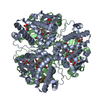
 集合体
集合体

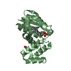


 分子量: 623.634 Da / 分子数: 2 / 由来タイプ: 合成 / 式: C35H33F4NO5
 分子量: 102.046 Da / 分子数: 3 / 由来タイプ: 合成 / 式: C3H2O4
 分子量: 18.015 Da / 分子数: 155 / 由来タイプ: 天然 / 式: H2O
 試料調製
試料調製 / ビームライン: BL9-2 / 波長: 0.97945 Å
 解析
解析