Mass: 40.078 Da / Num. of mol.: 4 / Source method: obtained synthetically / Formula: Ca
Sequence details
THIS DEPOSITED STRUCTURE OF HUMAN CALMODULIN WAS REFINED USING INDIVIDUAL DOMAIN COORDINATES OF ...THIS DEPOSITED STRUCTURE OF HUMAN CALMODULIN WAS REFINED USING INDIVIDUAL DOMAIN COORDINATES OF DEPOSITION 1MXE, WHICH CORRESPONDS TO THE CHICKEN CALMODULIN. THE SEQUENCES OF HUMAN AND CHICKEN CALMODULIN DIFFER IN TWO POSITIONS, 99 (TYR FOR HUMAN AND PHE FOR CHICKEN) AND 143 (GLN FOR HUMAN AND THR FOR CHICKEN). THEREFORE, THE SIDECHAIN COORDINATES OF RESIDUES 99 AND 143 IN THIS DEPOSITION DO NOT REPRESENT COMPOSITION OF THE SAMPLES USED FOR EXPERIMENTAL DATA COLLECTION. THIS DISCREPANCIES DO NOT PRODUCE ANY DIFFERENCES WHEN EXPERIMENTAL DATA ARE FITTED SINCE BACKBONE N-HN RDC AND CONTRAST-MATCHED SAXS DATA DO NOT INVOLVE THE SIDECHAIN COORDINATES AND THEIR IMPACT ON THE AQUEOUS SAXS DATA IS NEGLIGIBLE.
-
Experimental details
-
Experiment
Experiment
Method
Details (eV)
SOLUTION SCATTERING
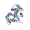
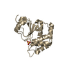
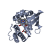
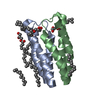
This entry contains the coordinates of the complex between Ca-bound human calmodulin and skeletal muscle myosin light chain kinase determined by refinement against backbone HN-N residual dipolar couplings from solution NMR and solution X-ray scattering intensity data. The latter include both SAXS data in H2O recorded on a Ca-CAM/MLCK sample and data in 65% aqueous sucrose buffer recorded on the Pb-CaM/MLCK sample in which Ca ions in calmodulin were replaced by Pb ions. The position of the C-terminal relative to the N-terminal domain of calmodulin was optimized via an exhaustive rigid-body translational/orientational grid search with a step size of 1A/2deg while fitting RDCs and the two types of SAXS data. Subsequently, the coordinates of the linker connecting the two domain of calmodulin (residues 76-81) and the interfacial side chains were optimized via molecular dynamics/simulated annealing refinement while keeping the coordinates of the backbone atoms fixed.
SOLUTION NMR
NMR experiment
Conditions-ID
Experiment-ID
Solution-ID
Type
1
1
1
2D 1H-15N HSQC
1
2
2
2D 1H-15N HSQC
-
Sample preparation
Details
Solution-ID
Contents
Solvent system
1
0.3 mM [U-13C; U-15N; U-2H] Calmodulin, 0.3 mM ssMLCK, 95% H2O/5% D2O
95% H2O/5% D2O
2
0.06-0.25 mM [U-13C; U-15N; U-2H] Calmodulin, 0.06-0.25 mM ssMLCK, 65% w/v sucrose/H2O
65% w/vsucrose/H2O
Sample
Conc. (mg/ml)
Units
Component
Isotopic labeling
Conc. range (mg/ml)
Solution-ID
0.3mM
Calmodulin-1
[U-13C; U-15N; U-2H]
1
0.3mM
ssMLCK-2
1
mM
Calmodulin-3
[U-13C; U-15N; U-2H]
0.06-0.25
2
mM
ssMLCK-4
0.06-0.25
2
Sample conditions
Conditions-ID
Ionic strength
pH
Pressure (kPa)
Temperature (K)
1
0.1
6.5
ambient
300K
2
0.15
6.5
ambient
300K
-
Data collection
NMR spectrometer
Type: Bruker DRX / Manufacturer: Bruker / Model: DRX / Field strength: 600 MHz
Soln scatter
Data reduction software list: MARDETECTOR, IGOR / Num. of time frames: 20 / Sample pH: 6.5 / Source class: Y / Source type: ADVANCED PHOTON SOURCE / Temperature: 298 K / Type: x-ray
Method: rigid-body optimization, simulated annealing / Software ordinal: 1 Details: Relative positions of the N- and C-terminal domains of calmodulin, rigidly held at those of the 1MXE structure, chain A, were optimized via a rotational/translational grid search with steps ...Details: Relative positions of the N- and C-terminal domains of calmodulin, rigidly held at those of the 1MXE structure, chain A, were optimized via a rotational/translational grid search with steps of 2deg and 1A, respectively with the best model fitted against RDC and scattering intensity data. Coordinates of the calmodulin linker residues (76-81) and the interfacial side chains were regularized via a molecular dynamics/simulated annealing protocol with the backbone atoms and the remaining side chains held fixed at the geometry resulting from the rigid-body grid search of the previous step.
NMR representative
Selection criteria: fewest violations
NMR ensemble
Conformer selection criteria: structures with the least restraint violations Conformers calculated total number: 1 / Conformers submitted total number: 1
Soln scatter model
Conformer selection criteria: BEST-FITTING CONFORMER BASED ON THE FIT OF AQUEOUS SAXS DATA FOR CA-CAM/MLCK, CONTRAST-MATCHED (65% SUCROSE) SAXS DATA FOR PB-CAM/MLCK, AND BACKBONE 1H-15N RESIDUAL ...Conformer selection criteria: BEST-FITTING CONFORMER BASED ON THE FIT OF AQUEOUS SAXS DATA FOR CA-CAM/MLCK, CONTRAST-MATCHED (65% SUCROSE) SAXS DATA FOR PB-CAM/MLCK, AND BACKBONE 1H-15N RESIDUAL DIPOLAR COUPLINGS FOR CAM IN THE CA-CAM/MLCK COMPLEX Details: 4000000000000 STARTING RELATIVE POSITIONS OF THE N-TERM (1-75) AND C-TERM (82-148) DOMAINS OF CAM WERE BUILT USING COORDINATES IN THE PDB DEPOSITION 1MXE:A. TAIL RESIDUES 1-4 AND 144-148 ...Details: 4000000000000 STARTING RELATIVE POSITIONS OF THE N-TERM (1-75) AND C-TERM (82-148) DOMAINS OF CAM WERE BUILT USING COORDINATES IN THE PDB DEPOSITION 1MXE:A. TAIL RESIDUES 1-4 AND 144-148 WERE BEST-FITTED TO 1MXE USING MODELS 1J7O AND 1J7P. THESE 4*1011 MODELS SAMPLE IN AN EXHAUSTIVE FASHION ALL POSSIBLE RELATIVE DOMAIN POSITIONS WHICH DIFFER BY NO MORE THAN 1A IN TERMS OF C-TERM DOMAIN PLACEMENT WHEN N-TERM DOMAIN IS FIXED. ROTATIONAL AND TRANSLATIONAL GRIDS OF 2DEG AND 1A WERE USED. THE MODELS WERE THEN FILTERED TO EXHIBIT FIT TO THE RDC DATA NO WORSE THAN 10% HIGHER THAN GLOBAL MINIMUM FOUND BY NLS OPTIMIZATION OF ORIENTATION OF THE C-TERM RELATIVE TO THE N-TERM DOMAIN. ADDITIONAL FILTERS WERE THEN APPLIED INCLUDING ABSENCE OF CLASHES <2.5 A BETWEEN HEAVY BACKBONE AND CB ATOMS, AND RGYR OF 13-19A. FOR THE GEOMETRIES THAT PASSED ABOVE REQUIREMENTS, POSITION OF MLCK FROM 2BBM DEPOSITION WAS BEST-FITTED IN A RIGID-BODY MANNER AGAINST NOE DISTANCE RESTRAINTS FROM 2BBM DEPOSITION AND CLASH AVOIDANCE FUNCTION INCLUDING HEAVY BACKBONE AND CB ATOMS. CAM CONFORMATIONS WHICH EXHIBITED AT LEAST 1 NOE VIOLATION EXCEEDING 3A FOR THE BEST-FITTED MLCK WERE REJECTED. LINKER COORDINATES WERE THEN BUILT FOR RESIDUES 76-81 USING 8-RESIDUE BACKBONE AND CB-CONTAINING SEGMENTS FROM A PDB-BASED DATABASE INCLUDING 2.2 MILLION RESIDUES IN TOTAL. LINKERS WHICH CLASHED WITH CAM DOMAINS OR MLCK OR EXHIBITED RMSD ABOVE 1.2 A FOR RESIDUES 1 AND 8 VS RESIDUES 75 AND 82 OF CAM WERE REJECTED. CAM CONFORMATION FOR WHICH SUCH LINKERS COULD NOT BE BUILT WERE REJECTED LEADING TO A TOTAL OF APPROXIMATELY 75000 STRUCTURES. THESE STRUCTURES WERE FITTED TO THE SAXS AND RDC DATA WITH THE OVERALL BEST-FITTING MODEL RETAINED FOR DEPOSITION. THE COORDINATES OF THE LINKER RESIDUES 76-81 AND SIDECHAINS WERE REGULARIZED BY ENERGY MINIMIZATION VIA XPLOR-NIH PRIOR TO THE DEPOSITION WITH THE ACTIVE ENERGY TERMS INCLUDING BONDS, ANGLES, IMPROPERS, NON-BONDED, AND CONFORMATIONAL DATABASE POTENTIALS OF MEAN FORCE. Num. of conformers submitted: 1 / Representative conformer: 1 / Software list: PRIMUS, GNOM
+
About Yorodumi
-
News
-
Feb 9, 2022. New format data for meta-information of EMDB entries
New format data for meta-information of EMDB entries
Version 3 of the EMDB header file is now the official format.
The previous official version 1.9 will be removed from the archive.
In the structure databanks used in Yorodumi, some data are registered as the other names, "COVID-19 virus" and "2019-nCoV". Here are the details of the virus and the list of structure data.
Jan 31, 2019. EMDB accession codes are about to change! (news from PDBe EMDB page)
EMDB accession codes are about to change! (news from PDBe EMDB page)
The allocation of 4 digits for EMDB accession codes will soon come to an end. Whilst these codes will remain in use, new EMDB accession codes will include an additional digit and will expand incrementally as the available range of codes is exhausted. The current 4-digit format prefixed with “EMD-” (i.e. EMD-XXXX) will advance to a 5-digit format (i.e. EMD-XXXXX), and so on. It is currently estimated that the 4-digit codes will be depleted around Spring 2019, at which point the 5-digit format will come into force.
The EM Navigator/Yorodumi systems omit the EMD- prefix.
Related info.:Q: What is EMD? / ID/Accession-code notation in Yorodumi/EM Navigator
Yorodumi is a browser for structure data from EMDB, PDB, SASBDB, etc.
This page is also the successor to EM Navigator detail page, and also detail information page/front-end page for Omokage search.
The word "yorodu" (or yorozu) is an old Japanese word meaning "ten thousand". "mi" (miru) is to see.
Related info.:EMDB / PDB / SASBDB / Comparison of 3 databanks / Yorodumi Search / Aug 31, 2016. New EM Navigator & Yorodumi / Yorodumi Papers / Jmol/JSmol / Function and homology information / Changes in new EM Navigator and Yorodumi
 Movie
Movie Controller

 Yorodumi
 Open data
 Basic information
 Components
 Keywords
 Function and homology information
 Homo sapiens (human)
 SOLUTION SCATTERING / SOLUTION NMR / rigid-body optimization, simulated annealing
  Authors
 Citation
 Structure visualization
 Downloads & links
 Other downloads
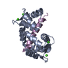


 PDBj
PDBj


 Assembly


 Mass: 40.078 Da / Num. of mol.: 4 / Source method: obtained synthetically / Formula: Ca
 Sample preparation
 Processing