Mass: 18.015 Da / Num. of mol.: 273 / Source method: isolated from a natural source / Formula: H2O
-
Details
Has protein modification
Y
-
Experimental details
-
Experiment
Experiment
Method: X-RAY DIFFRACTION / Number of used crystals: 1
-
Sample preparation
Crystal
Density Matthews: 2.02 Å3/Da / Density % sol: 39.06 %
Crystal grow
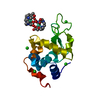
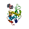
Temperature: 294 K / Method: vapor diffusion, hanging drop / pH: 4.5 Details: Tetragonal crystals were grown by the vapour-diffusion technique using the hanging-drop method, with 0.7-1.0 M sodium chloride and 50 mM sodium acetate buffer at pH 4.5. Derivative crystals ...Details: Tetragonal crystals were grown by the vapour-diffusion technique using the hanging-drop method, with 0.7-1.0 M sodium chloride and 50 mM sodium acetate buffer at pH 4.5. Derivative crystals containing gadolinium were obtained using concentrations of Gd-HPDO3A in the range 10-100 mM.
-
Data collection
Diffraction
ID
Mean temperature (K)
Crystal-ID
1
90
1
2
90
1
Diffraction source
Source
Site
Beamline
ID
Wavelength (Å)
SYNCHROTRON
ALS
8.3.1
1
0.97934
SYNCHROTRON
ALS
12.3.1
2
0.97934
Detector
Type
ID
Detector
Date
ADSC QUANTUM 315r
1
CCD
Jun 26, 2007
ADSC QUANTUM 315
2
CCD
Jun 25, 2007
Radiation
ID
Monochromator
Protocol
Monochromatic (M) / Laue (L)
Scattering type
Wavelength-ID
1
Si(111)
SINGLEWAVELENGTH
M
x-ray
1
2
Si(111)
SINGLEWAVELENGTH
M
x-ray
2
Radiation wavelength
ID
Wavelength (Å)
Relative weight
1
0.97934
1
2
1
Reflection
Resolution: 1.45→38.61 Å / Num. obs: 19784 / % possible obs: 87.8 % / Observed criterion σ(I): -3 / Redundancy: 3.4 % / Biso Wilson estimate: 11.64 Å2 / Rmerge(I) obs: 0.045 / Net I/σ(I): 17.57
Reflection shell
Resolution: 1.45→1.49 Å / Redundancy: 1.3 % / Rmerge(I) obs: 0.282 / Mean I/σ(I) obs: 1.86 / % possible all: 39
-
Processing
Software
Name
Version
Classification
PHENIX
1.9_1692
refinement
Blu-Ice
datacollection
XDS
datareduction
XSCALE
datascaling
PDB_EXTRACT
3.14
dataextraction
Refinement
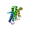
Method to determine structure: MOLECULAR REPLACEMENT Starting model: 1H87
In the structure databanks used in Yorodumi, some data are registered as the other names, "COVID-19 virus" and "2019-nCoV". Here are the details of the virus and the list of structure data.
Jan 31, 2019. EMDB accession codes are about to change! (news from PDBe EMDB page)
EMDB accession codes are about to change! (news from PDBe EMDB page)
The allocation of 4 digits for EMDB accession codes will soon come to an end. Whilst these codes will remain in use, new EMDB accession codes will include an additional digit and will expand incrementally as the available range of codes is exhausted. The current 4-digit format prefixed with “EMD-” (i.e. EMD-XXXX) will advance to a 5-digit format (i.e. EMD-XXXXX), and so on. It is currently estimated that the 4-digit codes will be depleted around Spring 2019, at which point the 5-digit format will come into force.
The EM Navigator/Yorodumi systems omit the EMD- prefix.
Related info.:Q: What is EMD? / ID/Accession-code notation in Yorodumi/EM Navigator
Yorodumi is a browser for structure data from EMDB, PDB, SASBDB, etc.
This page is also the successor to EM Navigator detail page, and also detail information page/front-end page for Omokage search.
The word "yorodu" (or yorozu) is an old Japanese word meaning "ten thousand". "mi" (miru) is to see.
Related info.:EMDB / PDB / SASBDB / Comparison of 3 databanks / Yorodumi Search / Aug 31, 2016. New EM Navigator & Yorodumi / Yorodumi Papers / Jmol/JSmol / Function and homology information / Changes in new EM Navigator and Yorodumi
 Movie
Movie Controller

 Yorodumi
 Open data
 Basic information
 Components
 Keywords
 Function and homology information
 X-RAY DIFFRACTION /
  Authors
 United States, 4items
  Citation
 Structure visualization
 Downloads & links
 Other downloads
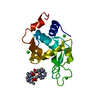


 PDBj
PDBj


 Assembly
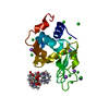


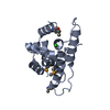
 Mass: 157.250 Da / Num. of mol.: 3 / Source method: obtained synthetically / Formula: Gd
 Mass: 404.459 Da / Num. of mol.: 3 / Source method: obtained synthetically / Formula: C17H32N4O7
 Mass: 35.453 Da / Num. of mol.: 13 / Source method: obtained synthetically / Formula: Cl
 Mass: 22.990 Da / Num. of mol.: 5 / Source method: isolated from a natural source / Formula: Na
 Sample preparation
 Processing