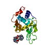
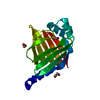
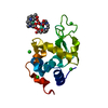
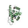
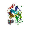
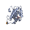
Entry Database : PDB / ID : 5oerTitle Hen egg-white lysozyme refined against 5000 9 keV diffraction patterns Lysozyme C Keywords / / / Function / homology Function Domain/homology Component
/ / / / / / / / / / / / / / / / / / / / / / / / / / / / / / / / / / / / / / / / / / / Biological species Gallus gallus (chicken)Method / / / Resolution : 1.9 Å Authors Gorel, A. / Schlichting, I. Journal : Nat Commun / Year : 2017Title : Multi-wavelength anomalous diffraction de novo phasing using a two-colour X-ray free-electron laser with wide tunability.Authors: Gorel, A. / Motomura, K. / Fukuzawa, H. / Doak, R.B. / Grunbein, M.L. / Hilpert, M. / Inoue, I. / Kloos, M. / Kovacsova, G. / Nango, E. / Nass, K. / Roome, C.M. / Shoeman, R.L. / Tanaka, R. ... Authors : Gorel, A. / Motomura, K. / Fukuzawa, H. / Doak, R.B. / Grunbein, M.L. / Hilpert, M. / Inoue, I. / Kloos, M. / Kovacsova, G. / Nango, E. / Nass, K. / Roome, C.M. / Shoeman, R.L. / Tanaka, R. / Tono, K. / Joti, Y. / Yabashi, M. / Iwata, S. / Foucar, L. / Ueda, K. / Barends, T.R.M. / Schlichting, I. History Deposition Jul 9, 2017 Deposition site / Processing site Revision 1.0 Oct 25, 2017 Provider / Type Revision 1.1 Nov 1, 2017 Group / Category Item _citation.country / _citation.journal_abbrev ... _citation.country / _citation.journal_abbrev / _citation.journal_id_CSD / _citation.journal_id_ISSN / _citation.pdbx_database_id_DOI / _citation.year Revision 1.2 Nov 8, 2017 Group / Category / citation_authorItem _citation.journal_volume / _citation.page_first ... _citation.journal_volume / _citation.page_first / _citation.page_last / _citation.pdbx_database_id_PubMed / _citation.title / _citation_author.name Revision 1.3 Jan 24, 2018 Group / Category Item / _diffrn_source.pdbx_synchrotron_siteRevision 1.4 Nov 14, 2018 Group / Category / Item Revision 1.5 Dec 13, 2023 Group / Database referencesCategory chem_comp_atom / chem_comp_bond ... chem_comp_atom / chem_comp_bond / database_2 / pdbx_related_exp_data_set Item / _database_2.pdbx_database_accessionRevision 1.6 Oct 23, 2024 Group / Category / pdbx_modification_feature
Show all Show less
 Movie
Movie Controller

 Yorodumi
 Open data
 Basic information
 Components
 Keywords
 Function and homology information
 X-RAY DIFFRACTION /
  Authors
 Citation
 Structure visualization
 Downloads & links
 Other downloads
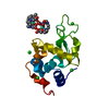


 PDBj
PDBj


 Assembly
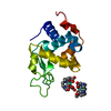
 Mass: 404.459 Da / Num. of mol.: 2 / Source method: obtained synthetically / Formula: C17H32N4O7
 Mass: 157.250 Da / Num. of mol.: 2 / Source method: obtained synthetically / Formula: Gd
 Mass: 22.990 Da / Num. of mol.: 1 / Source method: obtained synthetically / Formula: Na
 Mass: 18.015 Da / Num. of mol.: 70 / Source method: isolated from a natural source / Formula: H2O
 Sample preparation
 / Beamline: BL3 / Wavelength: 1.38 Å
 Processing